Terpyridine platinum(II) complexes inhibit cysteine proteases by binding to active-site cysteine.
Lo, Y.-C., Su, W.-C., Ko, T.-P., Wang, N.-C., Wang, A.H.-J.(2011) J Biomol Struct Dyn 29: 267-282
- PubMed: 21875148 Search on PubMed
- DOI: https://doi.org/10.1080/073911011010524993
- Primary Citation Related Structures:
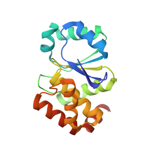
3RNZ, 3RO0, 3RO1 - PubMed Abstract:
Platinum(II) complexes have been demonstrated to form covalent bonds with sulfur-donating ligands (in glutathione, metallothionein and other sulfur-containing biomolecules) or coordination bonds with nitrogen-donating ligands (such as histidine and guanine). To investigate how these compounds interact with cysteine proteases, we chose terpyridine platinum(II) (TP-Pt(II)) complexes as a model system. By using X-ray crystallography, we demonstrated that TP-Pt(II) formed a covalent bond with the catalytic cysteine residue in pyroglutamyl peptidase I. Moreover, by using MALDI (matrix-assisted laser desorption/ionization) and TOF-TOF (time of flight) mass spectrometry, we elucidated that the TP-Pt(II) complex formed a covalent bond with the active-site cysteine residue in two other types of cysteine protease. Taken together, the results unequivocally showed that TP-Pt(II) complexes can selectively bind to the active site of most cysteine proteases. Our findings here can be useful in the design of new anti-cancer, anti-parasite or anti-virus platinum(II) compounds.
- Department and Institute of Pharmacology, National Yang-Ming University, Taipei 112, Taiwan.
Organizational Affiliation: