3RNY | pdb_00003rny

Crystal structure of human RSK1 C-terminal kinase domain


Experimental Data Snapshot

  • Method: X-RAY DIFFRACTION
  • Resolution: 2.70 Å
  • R-Value Free: 
    0.238 (Depositor), 0.258 (DCC) 
  • R-Value Work: 
    0.188 (Depositor), 0.207 (DCC) 
  • R-Value Observed: 
    0.191 (Depositor) 

Starting Model: experimental
View more details

wwPDB Validation 3D Report Full Report

Validation slider image for 3RNY

This is version 1.2 of the entry. See complete history

Literature

Structural basis for the autoinhibition of the C-terminal kinase domain of human RSK1.

Li, D.Fu, T.M.Nan, J.Liu, C.Li, L.F.Su, X.D.

(2012) Acta Crystallogr D Biol Crystallogr 68: 680-685

  • DOI: https://doi.org/10.1107/S0907444912007457
  • Primary Citation Related Structures: 
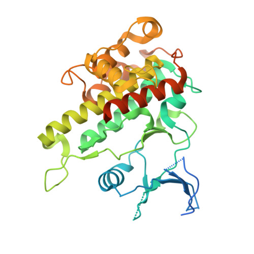
    3RNY

  • PubMed Abstract: 

    p90 ribosomal S6 kinases (RSKs) respond to various mitogen stimuli and comprise two distinct protein kinase domains. The C-terminal kinase domain (CTKD) receives signal from ERK1/2 and adopts an autoinhibitory mechanism. Here, the crystal structure of human RSK1 CTKD is reported at 2.7 Å resolution. The structure shows a standard kinase fold, with the catalytic residues in the ATP-binding cleft orientated in optimal conformations for phosphotransfer. The inactivation of the CTKD is conferred by an extra α-helix (αL), which occupies the substrate-binding groove. In combination with previous knowledge, this structure indicates that activation of RSK1 involves the removal of αL from the substrate-binding groove induced by ERK1/2 phosphorylation.


  • Organizational Affiliation
    • State Key Laboratory of Protein and Plant Gene Research, School of Life Sciences, Peking University, Beijing 100871, People's Republic of China.

Macromolecule Content 

  • Total Structure Weight: 77.69 kDa 
  • Atom Count: 4,068 
  • Modeled Residue Count: 535 
  • Deposited Residue Count: 692 
  • Unique protein chains: 1

Macromolecules

Find similar proteins by:|  3D Structure
Entity ID: 1
MoleculeChains  Sequence LengthOrganismDetailsImage
Ribosomal protein S6 kinase alpha-1
A, B
346Homo sapiensMutation(s): 0 
Gene Names: RPS6KA1MAPKAPK1ARSK1
EC: 2.7.11.1
UniProt & NIH Common Fund Data Resources
Find proteins for Q15418 (Homo sapiens)
Explore Q15418 
Go to UniProtKB:  Q15418
PHAROS:  Q15418
GTEx:  ENSG00000117676 
Entity Groups
Sequence Clusters30% Identity50% Identity70% Identity90% Identity95% Identity100% Identity
UniProt GroupQ15418
Sequence Annotations
Expand
Reference Sequence

Experimental Data & Validation

Experimental Data

  • Method: X-RAY DIFFRACTION
  • Resolution: 2.70 Å
  • R-Value Free:  0.238 (Depositor), 0.258 (DCC) 
  • R-Value Work:  0.188 (Depositor), 0.207 (DCC) 
  • R-Value Observed: 0.191 (Depositor) 
Space Group: P 1 21 1
Unit Cell:
Length ( Å )Angle ( ˚ )
a = 39.86α = 90
b = 143.47β = 95.77
c = 59.92γ = 90
Software Package:
Software NamePurpose
BUSTER-TNTrefinement
PDB_EXTRACTdata extraction
HKL-2000data collection
HKL-2000data reduction
HKL-2000data scaling
MOLREPphasing
BUSTERrefinement

Structure Validation

View Full Validation Report


Entry History 

Deposition Data

Revision History  (Full details and data files)

  • Version 1.0: 2012-04-25
    Type: Initial release
  • Version 1.1: 2012-08-29
    Changes: Database references
  • Version 1.2: 2023-09-13
    Changes: Data collection, Database references, Derived calculations, Refinement description