3RLJ | pdb_00003rlj

Crystal structure of the androgen receptor ligand binding domain in complex with SARM S-22


Experimental Data Snapshot

  • Method: X-RAY DIFFRACTION
  • Resolution: 1.90 Å
  • R-Value Free: 
    0.265 (Depositor), 0.259 (DCC) 
  • R-Value Work: 
    0.221 (Depositor), 0.214 (DCC) 
  • R-Value Observed: 
    0.221 (Depositor) 

Starting Model: experimental
View more details

wwPDB Validation 3D Report Full Report

Validation slider image for 3RLJ

Ligand Structure Quality Assessment 


This is version 1.2 of the entry. See complete history

Literature

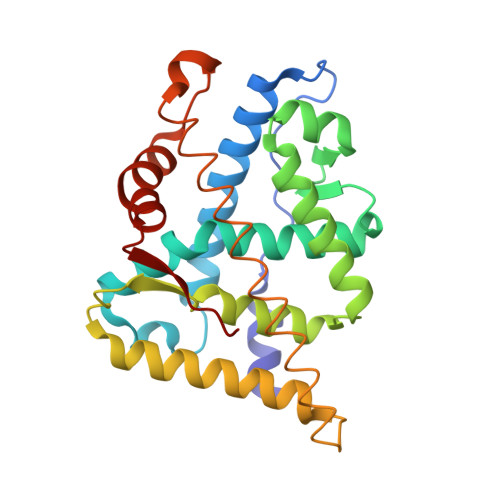
Unexpected binding orientation of bulky-B-ring anti-androgens and implications for future drug targets.

Duke, C.B.Jones, A.Bohl, C.E.Dalton, J.T.Miller, D.D.

(2011) J Med Chem 54: 3973-3976

  • DOI: https://doi.org/10.1021/jm2000097
  • Primary Citation Related Structures: 
    3RLJ, 3RLL

  • PubMed Abstract: 

    Several new androgen receptor antagonists were synthesized and found to have varying activities across typically anti-androgen resistant mutants (Thr877 → Ala and Trp741 → Leu) and markedly improved potency over previously reported pan-antagonists. X-ray crystallography of a new anti-androgen in an androgen receptor mutant (Thr877 → Ala) shows that the receptor can accommodate the added bulk presented by phenyl to naphthyl substitution, casting doubt on previous reports of predicted binding orientation and the causes of antagonism in bulky-B-ring antagonists.


  • Organizational Affiliation
    • Department of Pharmaceutical Sciences, The University of Tennessee, Memphis, TN 38163, USA.

Macromolecule Content 

  • Total Structure Weight: 29.21 kDa 
  • Atom Count: 2,225 
  • Modeled Residue Count: 247 
  • Deposited Residue Count: 247 
  • Unique protein chains: 1

Macromolecules

Find similar proteins by:|  3D Structure
Entity ID: 1
MoleculeChains  Sequence LengthOrganismDetailsImage
Androgen receptor247Homo sapiensMutation(s): 0 
Gene Names: ARDHTRNR3C4
UniProt & NIH Common Fund Data Resources
Find proteins for P10275 (Homo sapiens)
Explore P10275 
Go to UniProtKB:  P10275
PHAROS:  P10275
GTEx:  ENSG00000169083 
Entity Groups
Sequence Clusters30% Identity50% Identity70% Identity90% Identity95% Identity100% Identity
UniProt GroupP10275
Sequence Annotations
Expand
Reference Sequence

Small Molecules

Ligands 1 Unique
IDChains Name / Formula / InChI Key2D Diagram3D Interactions
RLJ

Query on RLJ


Download:Ideal Coordinates CCD File
B [auth A](2S)-3-(4-cyanophenoxy)-N-[4-cyano-3-(trifluoromethyl)phenyl]-2-hydroxy-2-methylpropanamide
C19 H14 F3 N3 O3
JNGVJMBLXIUVRD-SFHVURJKSA-N
Binding Affinity Annotations 
IDSourceBinding Affinity
RLJ BindingDB:  3RLJ IC50: 36 (nM) from 1 assay(s)

Experimental Data & Validation

Experimental Data

  • Method: X-RAY DIFFRACTION
  • Resolution: 1.90 Å
  • R-Value Free:  0.265 (Depositor), 0.259 (DCC) 
  • R-Value Work:  0.221 (Depositor), 0.214 (DCC) 
  • R-Value Observed: 0.221 (Depositor) 
Space Group: P 21 21 21
Unit Cell:
Length ( Å )Angle ( ˚ )
a = 54.742α = 90
b = 66.795β = 90
c = 69.374γ = 90
Software Package:
Software NamePurpose
CrystalCleardata collection
CNSrefinement
d*TREKdata reduction
d*TREKdata scaling
CNSphasing

Structure Validation

View Full Validation Report


Ligand Structure Quality Assessment 


Entry History 

Deposition Data

Revision History  (Full details and data files)

  • Version 1.0: 2011-05-04
    Type: Initial release
  • Version 1.1: 2011-07-13
    Changes: Version format compliance
  • Version 1.2: 2023-09-13
    Changes: Data collection, Database references, Derived calculations, Refinement description