3RLE | pdb_00003rle

Crystal Structure of GRASP55 GRASP domain (residues 7-208)


Experimental Data Snapshot

  • Method: X-RAY DIFFRACTION
  • Resolution: 1.65 Å
  • R-Value Free: 
    0.222 (Depositor), 0.227 (DCC) 
  • R-Value Work: 
    0.176 (Depositor), 0.183 (DCC) 
  • R-Value Observed: 
    0.179 (Depositor) 

wwPDB Validation 3D Report Full Report

Validation slider image for 3RLE

This is version 1.2 of the entry. See complete history

Literature

Structure of the Membrane-tethering GRASP Domain Reveals a Unique PDZ Ligand Interaction That Mediates Golgi Biogenesis.

Truschel, S.T.Sengupta, D.Foote, A.Heroux, A.Macbeth, M.R.Linstedt, A.D.

(2011) J Biological Chem 286: 20125-20129

  • DOI: https://doi.org/10.1074/jbc.C111.245324
  • Primary Citation Related Structures: 
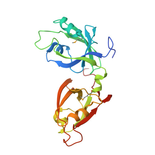
    3RLE

  • PubMed Abstract: 

    Biogenesis of the ribbon-like membrane network of the mammalian Golgi requires membrane tethering by the conserved GRASP domain in GRASP65 and GRASP55, yet the tethering mechanism is not fully understood. Here, we report the crystal structure of the GRASP55 GRASP domain, which revealed an unusual arrangement of two tandem PDZ folds that more closely resemble prokaryotic PDZ domains. Biochemical and functional data indicated that the interaction between the ligand-binding pocket of PDZ1 and an internal ligand on PDZ2 mediates the GRASP self-interaction, and structural analyses suggest that this occurs via a unique mode of internal PDZ ligand recognition. Our data uncover the structural basis for ligand specificity and provide insight into the mechanism of GRASP-dependent membrane tethering of analogous Golgi cisternae.


  • Organizational Affiliation
    • Department of Biological Sciences, Carnegie Mellon University, Pittsburgh, Pennsylvania 15213, USA.

Macromolecule Content 

  • Total Structure Weight: 22.99 kDa 
  • Atom Count: 1,842 
  • Modeled Residue Count: 202 
  • Deposited Residue Count: 209 
  • Unique protein chains: 1

Macromolecules

Find similar proteins by:|  3D Structure
Entity ID: 1
MoleculeChains  Sequence LengthOrganismDetailsImage
Golgi reassembly-stacking protein 2209Homo sapiensMutation(s): 0 
Gene Names: GORASP2GOLPH6
UniProt & NIH Common Fund Data Resources
Find proteins for Q9H8Y8 (Homo sapiens)
Explore Q9H8Y8 
Go to UniProtKB:  Q9H8Y8
PHAROS:  Q9H8Y8
GTEx:  ENSG00000115806 
Entity Groups
Sequence Clusters30% Identity50% Identity70% Identity90% Identity95% Identity100% Identity
UniProt GroupQ9H8Y8
Sequence Annotations
Expand
Reference Sequence

Small Molecules

Modified Residues  1 Unique
IDChains TypeFormula2D DiagramParent
MSE
Query on MSE
A
L-PEPTIDE LINKINGC5 H11 N O2 SeMET

Experimental Data & Validation

Experimental Data

  • Method: X-RAY DIFFRACTION
  • Resolution: 1.65 Å
  • R-Value Free:  0.222 (Depositor), 0.227 (DCC) 
  • R-Value Work:  0.176 (Depositor), 0.183 (DCC) 
  • R-Value Observed: 0.179 (Depositor) 
Space Group: P 41 21 2
Unit Cell:
Length ( Å )Angle ( ˚ )
a = 88.04α = 90
b = 88.04β = 90
c = 61.195γ = 90
Software Package:
Software NamePurpose
HKL-2000data collection
SHELXSphasing
REFMACrefinement
HKL-2000data reduction
HKL-2000data scaling

Structure Validation

View Full Validation Report


Entry History 

Deposition Data

Revision History  (Full details and data files)

  • Version 1.0: 2011-05-04
    Type: Initial release
  • Version 1.1: 2011-07-13
    Changes: Version format compliance
  • Version 1.2: 2024-10-09
    Changes: Data collection, Database references, Derived calculations, Structure summary