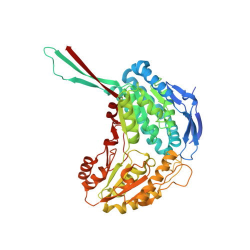
3RHJ

Crystal structure of the E673A mutant of the C-terminal domain of RAT 10'FORMYLTETRAHYDROFOLATE DEHYDROGENASE in complex with co-purified NADP


Experimental Data Snapshot

  • Method: X-RAY DIFFRACTION
  • Resolution: 1.89 Å
  • R-Value Free: 0.203 
  • R-Value Work: 0.189 
  • R-Value Observed: 0.189 

Starting Model: experimental
View more details

wwPDB Validation   3D Report Full Report


Ligand Structure Quality Assessment 


This is version 1.3 of the entry. See complete history


Literature

Conserved catalytic residues of the ALDH1L1 aldehyde dehydrogenase domain control binding and discharging of the coenzyme.

Tsybovsky, Y.Krupenko, S.A.

(2011) J Biol Chem 286: 23357-23367

  • DOI: https://doi.org/10.1074/jbc.M111.221069
  • Primary Citation of Related Structures:  
    3RHJ, 3RHL, 3RHM, 3RHO, 3RHP, 3RHQ, 3RHR

  • PubMed Abstract: 

    The C-terminal domain (C(t)-FDH) of 10-formyltetrahydrofolate dehydrogenase (FDH, ALDH1L1) is an NADP(+)-dependent oxidoreductase and a structural and functional homolog of aldehyde dehydrogenases. Here we report the crystal structures of several C(t)-FDH mutants in which two essential catalytic residues adjacent to the nicotinamide ring of bound NADP(+), Cys-707 and Glu-673, were replaced separately or simultaneously. The replacement of the glutamate with an alanine causes irreversible binding of the coenzyme without any noticeable conformational changes in the vicinity of the nicotinamide ring. Additional replacement of cysteine 707 with an alanine (E673A/C707A double mutant) did not affect this irreversible binding indicating that the lack of the glutamate is solely responsible for the enhanced interaction between the enzyme and the coenzyme. The substitution of the cysteine with an alanine did not affect binding of NADP(+) but resulted in the enzyme lacking the ability to differentiate between the oxidized and reduced coenzyme: unlike the wild-type C(t)-FDH/NADPH complex, in the C707A mutant the position of NADPH is identical to the position of NADP(+) with the nicotinamide ring well ordered within the catalytic center. Thus, whereas the glutamate restricts the affinity for the coenzyme, the cysteine is the sensor of the coenzyme redox state. These conclusions were confirmed by coenzyme binding experiments. Our study further suggests that the binding of the coenzyme is additionally controlled by a long-range communication between the catalytic center and the coenzyme-binding domain and points toward an α-helix involved in the adenine moiety binding as a participant of this communication.


  • Organizational Affiliation

    Department of Biochemistry and Molecular Biology, Medical University of South Carolina, Charleston, South Carolina 29425, USA.


Macromolecules
Find similar proteins by:  (by identity cutoff)  |  3D Structure
Entity ID: 1
MoleculeChains Sequence LengthOrganismDetailsImage
Aldehyde dehydrogenase 1 family, member L1
A, B, C, D
517Rattus norvegicusMutation(s): 1 
Gene Names: Aldh1l1Fthfd
UniProt
Find proteins for P28037 (Rattus norvegicus)
Explore P28037 
Go to UniProtKB:  P28037
Entity Groups  
Sequence Clusters30% Identity50% Identity70% Identity90% Identity95% Identity100% Identity
UniProt GroupP28037
Sequence Annotations
Expand
  • Reference Sequence
Small Molecules
Ligands 3 Unique
IDChains Name / Formula / InChI Key2D Diagram3D Interactions
NAP
Query on NAP

Download Ideal Coordinates CCD File 
DA [auth D],
E [auth A],
M [auth B],
U [auth C]
NADP NICOTINAMIDE-ADENINE-DINUCLEOTIDE PHOSPHATE
C21 H28 N7 O17 P3
XJLXINKUBYWONI-NNYOXOHSSA-N
SO4
Query on SO4

Download Ideal Coordinates CCD File 
AA [auth C]
BA [auth C]
EA [auth D]
F [auth A]
FA [auth D]
AA [auth C],
BA [auth C],
EA [auth D],
F [auth A],
FA [auth D],
G [auth A],
GA [auth D],
H [auth A],
HA [auth D],
I [auth A],
IA [auth D],
J [auth A],
K [auth A],
N [auth B],
O [auth B],
P [auth B],
Q [auth B],
R [auth B],
S [auth B],
V [auth C],
W [auth C],
X [auth C],
Y [auth C],
Z [auth C]
SULFATE ION
O4 S
QAOWNCQODCNURD-UHFFFAOYSA-L
GOL
Query on GOL

Download Ideal Coordinates CCD File 
CA [auth C],
JA [auth D],
L [auth A],
T [auth B]
GLYCEROL
C3 H8 O3
PEDCQBHIVMGVHV-UHFFFAOYSA-N
Experimental Data & Validation

Experimental Data

  • Method: X-RAY DIFFRACTION
  • Resolution: 1.89 Å
  • R-Value Free: 0.203 
  • R-Value Work: 0.189 
  • R-Value Observed: 0.189 
  • Space Group: C 1 2 1
Unit Cell:
Length ( Å )Angle ( ˚ )
a = 259.391α = 90
b = 194.933β = 108.94
c = 97.48γ = 90
Software Package:
Software NamePurpose
SERGUIdata collection
MOLREPphasing
REFMACrefinement
HKL-2000data reduction
HKL-2000data scaling

Structure Validation

View Full Validation Report


Ligand Structure Quality Assessment 


Entry History 

Deposition Data

Revision History  (Full details and data files)

  • Version 1.0: 2011-04-27
    Type: Initial release
  • Version 1.1: 2011-07-13
    Changes: Version format compliance
  • Version 1.2: 2011-08-31
    Changes: Database references
  • Version 1.3: 2023-09-13
    Changes: Data collection, Database references, Derived calculations, Refinement description