3RGS

Structural and kinetic analysis of the beef liver catalase with the ammonia as a ligand


Experimental Data Snapshot

  • Method: X-RAY DIFFRACTION
  • Resolution: 1.99 Å
  • R-Value Free: 0.198 
  • R-Value Work: 0.161 
  • R-Value Observed: 0.161 

wwPDB Validation   3D Report Full Report


Ligand Structure Quality Assessment 


This is version 1.3 of the entry. See complete history


Literature

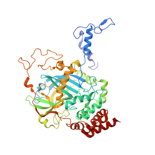
Interaction of nitric oxide with catalase: structural and kinetic analysis.

Purwar, N.McGarry, J.M.Kostera, J.Pacheco, A.A.Schmidt, M.

(2011) Biochemistry 50: 4491-4503

  • DOI: https://doi.org/10.1021/bi200130r
  • Primary Citation of Related Structures:  
    3RE8, 3RGP, 3RGS

  • PubMed Abstract: 

    We present the structures of bovine catalase in its native form and complexed with ammonia and nitric oxide, obtained by X-ray crystallography. Using the NO generator 1-(N,N-diethylamino)diazen-1-ium-1,2-diolate, we were able to generate sufficiently high NO concentrations within the catalase crystals that substantial occupation was observed despite a high dissociation rate. Nitric oxide seems to be slightly bent from the heme normal that may indicate some iron(II) character in the formally ferric catalase. Microspectrophotometric investigations inline with the synchrotron X-ray beam reveal photoreduction of the central heme iron. In the cases of the native and ammonia-complexed catalase, reduction is accompanied by a relaxation phase. This is likely not the case for the catalase NO complex. The kinetics of binding of NO to catalase were investigated using NO photolyzed from N,N'-bis(carboxymethyl)-N,N'-dinitroso-p-phenylenediamine using an assay that combines catalase with myoglobin binding kinetics. The off rate is 1.5 s(-1). Implications for catalase function are discussed.


  • Organizational Affiliation

    Department of Physics, University of Wisconsin-Milwaukee, 1900 East Kenwood Boulevard, Milwaukee, WI 53211, USA.


Macromolecules
Find similar proteins by:  (by identity cutoff)  |  3D Structure
Entity ID: 1
MoleculeChains Sequence LengthOrganismDetailsImage
Catalase
A, B, C, D
499Bos taurusMutation(s): 0 
EC: 1.11.1.6
UniProt
Find proteins for P00432 (Bos taurus)
Explore P00432 
Go to UniProtKB:  P00432
Entity Groups  
Sequence Clusters30% Identity50% Identity70% Identity90% Identity95% Identity100% Identity
UniProt GroupP00432
Sequence Annotations
Expand
  • Reference Sequence
Experimental Data & Validation

Experimental Data

  • Method: X-RAY DIFFRACTION
  • Resolution: 1.99 Å
  • R-Value Free: 0.198 
  • R-Value Work: 0.161 
  • R-Value Observed: 0.161 
  • Space Group: P 21 21 21
Unit Cell:
Length ( Å )Angle ( ˚ )
a = 83.48α = 90
b = 140.72β = 90
c = 229.52γ = 90
Software Package:
Software NamePurpose
HKL-3000data collection
CNSrefinement
MOSFLMdata reduction
SCALAdata scaling
CNSphasing

Structure Validation

View Full Validation Report


Ligand Structure Quality Assessment 


Entry History 

Deposition Data

Revision History  (Full details and data files)

  • Version 1.0: 2011-05-11
    Type: Initial release
  • Version 1.1: 2011-07-13
    Changes: Version format compliance
  • Version 1.2: 2012-04-04
    Changes: Database references
  • Version 1.3: 2023-09-13
    Changes: Data collection, Database references, Derived calculations, Refinement description