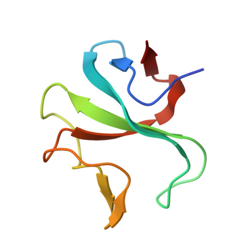
SLAIN2 links microtubule plus end-tracking proteins and controls microtubule growth in interphase
van der Vaart, B., Manatschal, C., Grigoriev, I., Olieric, V., Gouveia, S.M., Bjelic, S., Demmers, J., Vorobjev, I., Hoogenraad, C.C., Steinmetz, M.O., Akhmanova, A.(2011) J Cell Biol 193: 1083-1099
- PubMed: 21646404 Search on PubMedSearch on PubMed Central
- DOI: https://doi.org/10.1083/jcb.201012179
- Primary Citation Related Structures:
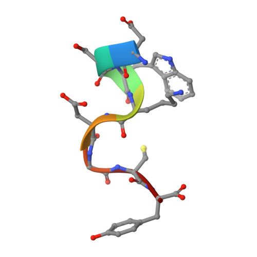
3RDV - PubMed Abstract:
The ends of growing microtubules (MTs) accumulate a set of diverse factors known as MT plus end-tracking proteins (+TIPs), which control microtubule dynamics and organization. In this paper, we identify SLAIN2 as a key component of +TIP interaction networks. We showed that the C-terminal part of SLAIN2 bound to end-binding proteins (EBs), cytoplasmic linker proteins (CLIPs), and CLIP-associated proteins and characterized in detail the interaction of SLAIN2 with EB1 and CLIP-170. Furthermore, we found that the N-terminal part of SLAIN2 interacted with ch-TOG, the mammalian homologue of the MT polymerase XMAP215. Through its multiple interactions, SLAIN2 enhanced ch-TOG accumulation at MT plus ends and, as a consequence, strongly stimulated processive MT polymerization in interphase cells. Depletion or disruption of the SLAIN2-ch-TOG complex led to disorganization of the radial MT array. During mitosis, SLAIN2 became highly phosphorylated, and its interaction with EBs and ch-TOG was inhibited. Our study provides new insights into the molecular mechanisms underlying cell cycle-specific regulation of MT polymerization and the organization of the MT network.
- Department of Cell Biology, Erasmus Medical Center, 3000 CA Rotterdam, Netherlands.
Organizational Affiliation: