3RC4 | pdb_00003rc4

Molecular mechanisms of viral and host-cell substrate recognition by HCV NS3/4A protease


Experimental Data Snapshot

  • Method: X-RAY DIFFRACTION
  • Resolution: 1.50 Å
  • R-Value Free: 
    0.215 (Depositor), 0.230 (DCC) 
  • R-Value Work: 
    0.186 (Depositor), 0.195 (DCC) 
  • R-Value Observed: 
    0.188 (Depositor) 

Starting Model: experimental
View more details

wwPDB Validation 3D Report Full Report

Validation slider image for 3RC4

This is version 1.5 of the entry. See complete history

Literature

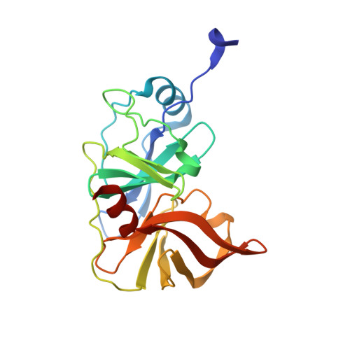
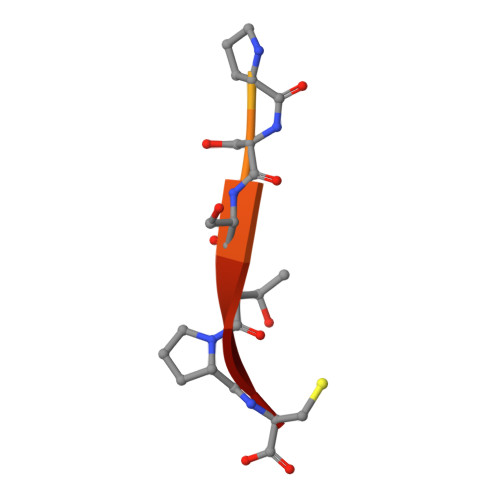
Molecular mechanisms of viral and host cell substrate recognition by hepatitis C virus NS3/4A protease.

Romano, K.P.Laine, J.M.Deveau, L.M.Cao, H.Massi, F.Schiffer, C.A.

(2011) J Virol 85: 6106-6116

  • DOI: https://doi.org/10.1128/JVI.00377-11
  • Primary Citation Related Structures: 
    3RC4, 3RC5, 3RC6

  • PubMed Abstract: 

    Hepatitis C NS3/4A protease is a prime therapeutic target that is responsible for cleaving the viral polyprotein at junctions 3-4A, 4A4B, 4B5A, and 5A5B and two host cell adaptor proteins of the innate immune response, TRIF and MAVS. In this study, NS3/4A crystal structures of both host cell cleavage sites were determined and compared to the crystal structures of viral substrates. Two distinct protease conformations were observed and correlated with substrate specificity: (i) 3-4A, 4A4B, 5A5B, and MAVS, which are processed more efficiently by the protease, form extensive electrostatic networks when in complex with the protease, and (ii) TRIF and 4B5A, which contain polyproline motifs in their full-length sequences, do not form electrostatic networks in their crystal complexes. These findings provide mechanistic insights into NS3/4A substrate recognition, which may assist in a more rational approach to inhibitor design in the face of the rapid acquisition of resistance.


  • Organizational Affiliation
    • University of Massachusetts Medical School, Department of Biochemistry and Molecular Pharmacology, 364 Plantation Street, Worcester, MA 01605, USA.

Macromolecule Content 

  • Total Structure Weight: 22.92 kDa 
  • Atom Count: 1,645 
  • Modeled Residue Count: 205 
  • Deposited Residue Count: 216 
  • Unique protein chains: 2

Macromolecules

Find similar proteins by:|  3D Structure
Entity ID: 1
MoleculeChains  Sequence LengthOrganismDetailsImage
NS3/4A Protease203hepatitis C virus genotype 1aMutation(s): 16 
UniProt
Find proteins for P26664 (Hepatitis C virus genotype 1a (isolate 1))
Explore P26664 
Go to UniProtKB:  P26664
Entity Groups
Sequence Clusters30% Identity50% Identity70% Identity90% Identity95% Identity100% Identity
UniProt GroupP26664
Sequence Annotations
Expand
Reference Sequence
Find similar proteins by:  Sequence   |   3D Structure  
Entity ID: 2
MoleculeChains  Sequence LengthOrganismDetailsImage
Product TRIF13Homo sapiensMutation(s): 0 
UniProt & NIH Common Fund Data Resources
Find proteins for Q8IUC6 (Homo sapiens)
Explore Q8IUC6 
Go to UniProtKB:  Q8IUC6
PHAROS:  Q8IUC6
GTEx:  ENSG00000127666 
Entity Groups
Sequence Clusters30% Identity50% Identity70% Identity90% Identity95% Identity100% Identity
UniProt GroupQ8IUC6
Sequence Annotations
Expand
Reference Sequence

Experimental Data & Validation

Experimental Data

  • Method: X-RAY DIFFRACTION
  • Resolution: 1.50 Å
  • R-Value Free:  0.215 (Depositor), 0.230 (DCC) 
  • R-Value Work:  0.186 (Depositor), 0.195 (DCC) 
  • R-Value Observed: 0.188 (Depositor) 
Space Group: P 21 21 21
Unit Cell:
Length ( Å )Angle ( ˚ )
a = 53.9α = 90
b = 58.146β = 90
c = 61.3γ = 90
Software Package:
Software NamePurpose
DENZOdata reduction
SCALEPACKdata scaling
REFMACrefinement
PDB_EXTRACTdata extraction

Structure Validation

View Full Validation Report


Entry History 

Deposition Data

Revision History  (Full details and data files)

  • Version 1.0: 2011-05-04
    Type: Initial release
  • Version 1.1: 2011-07-13
    Changes: Version format compliance
  • Version 1.2: 2017-06-28
    Changes: Database references, Source and taxonomy, Structure summary
  • Version 1.3: 2017-11-08
    Changes: Refinement description
  • Version 1.4: 2023-07-26
    Changes: Database references, Derived calculations
  • Version 1.5: 2023-09-13
    Changes: Data collection, Refinement description