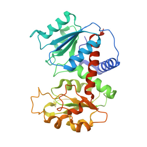
3R7F

Crystal Structure of CP-bound Aspartate Transcarbamoylase from Bacillus subtilis


Experimental Data Snapshot

  • Method: X-RAY DIFFRACTION
  • Resolution: 2.10 Å
  • R-Value Free: 0.195 
  • R-Value Work: 0.171 
  • R-Value Observed: 0.171 

wwPDB Validation   3D Report Full Report


This is version 1.4 of the entry. See complete history


Literature

Crystallographic Snapshots of the Complete Catalytic Cycle of the Unregulated Aspartate Transcarbamoylase from Bacillus subtilis.

Harris, K.M.Cockrell, G.M.Puleo, D.E.Kantrowitz, E.R.

(2011) J Mol Biol 411: 190-200

  • DOI: https://doi.org/10.1016/j.jmb.2011.05.036
  • Primary Citation of Related Structures:  
    3R7D, 3R7F, 3R7L

  • PubMed Abstract: 

    Here, we report high-resolution X-ray structures of Bacillus subtilis aspartate transcarbamoylase (ATCase), an enzyme that catalyzes one of the first reactions in pyrimidine nucleotide biosynthesis. Structures of the enzyme have been determined in the absence of ligands, in the presence of the substrate carbamoyl phosphate, and in the presence of the bisubstrate/transition state analog N-phosphonacetyl-L-aspartate. Combining the structural data with in silico docking and electrostatic calculations, we have been able to visualize each step in the catalytic cycle of ATCase, from the ordered binding of the substrates, to the formation and decomposition of the tetrahedral intermediate, to the ordered release of the products from the active site. Analysis of the conformational changes associated with these steps provides a rationale for the lack of cooperativity in trimeric ATCases that do not possess regulatory subunits.


  • Organizational Affiliation

    Department of Chemistry, Boston College, Merkert Chemistry Center, Chestnut Hill, MA 02467, USA.


Macromolecules
Find similar proteins by:  (by identity cutoff)  |  3D Structure
Entity ID: 1
MoleculeChains Sequence LengthOrganismDetailsImage
Aspartate carbamoyltransferase
A, B, C
304Bacillus subtilisMutation(s): 0 
Gene Names: pyrBBSU15490
EC: 2.1.3.2
UniProt
Find proteins for P05654 (Bacillus subtilis (strain 168))
Explore P05654 
Go to UniProtKB:  P05654
Entity Groups  
Sequence Clusters30% Identity50% Identity70% Identity90% Identity95% Identity100% Identity
UniProt GroupP05654
Sequence Annotations
Expand
  • Reference Sequence
Small Molecules
Ligands 2 Unique
IDChains Name / Formula / InChI Key2D Diagram3D Interactions
CP
Query on CP

Download Ideal Coordinates CCD File 
D [auth A],
K [auth B],
O [auth B],
P [auth C]
PHOSPHORIC ACID MONO(FORMAMIDE)ESTER
C H4 N O5 P
FFQKYPRQEYGKAF-UHFFFAOYSA-N
PO4
Query on PO4

Download Ideal Coordinates CCD File 
E [auth A]
F [auth A]
G [auth A]
H [auth A]
I [auth A]
E [auth A],
F [auth A],
G [auth A],
H [auth A],
I [auth A],
J [auth A],
L [auth B],
M [auth B],
N [auth B],
Q [auth C],
R [auth C],
S [auth C],
T [auth C]
PHOSPHATE ION
O4 P
NBIIXXVUZAFLBC-UHFFFAOYSA-K
Experimental Data & Validation

Experimental Data

  • Method: X-RAY DIFFRACTION
  • Resolution: 2.10 Å
  • R-Value Free: 0.195 
  • R-Value Work: 0.171 
  • R-Value Observed: 0.171 
  • Space Group: C 1 2 1
Unit Cell:
Length ( Å )Angle ( ˚ )
a = 251.916α = 90
b = 152.96β = 96.1
c = 51.239γ = 90
Software Package:
Software NamePurpose
DENZOdata reduction
SCALEPACKdata scaling
PHENIXrefinement
PDB_EXTRACTdata extraction
HKL-2000data reduction
HKL-2000data scaling

Structure Validation

View Full Validation Report


Entry History 

Deposition Data

Revision History  (Full details and data files)

  • Version 1.0: 2011-06-08
    Type: Initial release
  • Version 1.1: 2011-07-13
    Changes: Version format compliance
  • Version 1.2: 2011-08-10
    Changes: Database references
  • Version 1.3: 2017-11-08
    Changes: Advisory, Refinement description
  • Version 1.4: 2024-02-21
    Changes: Advisory, Data collection, Database references, Derived calculations