3R6R

Structure of the complex of an intramolecular human telomeric DNA with Berberine formed in K+ solution


Experimental Data Snapshot

  • Method: X-RAY DIFFRACTION
  • Resolution: 2.40 Å
  • R-Value Free: 0.252 
  • R-Value Work: 0.215 
  • R-Value Observed: 0.217 

wwPDB Validation   3D Report Full Report


Ligand Structure Quality Assessment 


This is version 1.2 of the entry. See complete history


Literature

The crystal structure of human telomeric DNA complexed with berberine: an interesting case of stacked ligand to G-tetrad ratio higher than 1:1.

Bazzicalupi, C.Ferraroni, M.Bilia, A.R.Scheggi, F.Gratteri, P.

(2013) Nucleic Acids Res 41: 632-638

  • DOI: https://doi.org/10.1093/nar/gks1001
  • Primary Citation of Related Structures:  
    3R6R

  • PubMed Abstract: 

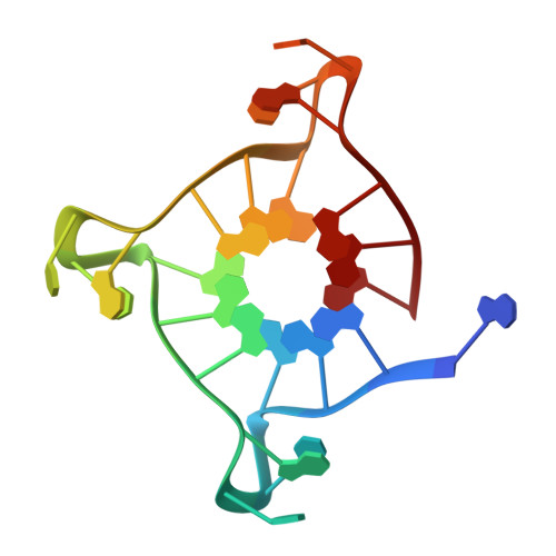
    The first crystal structure of human telomeric DNA in complex with the natural alkaloid berberine, produced by different plant families and used in folk medicine for millennia, was solved by X-ray diffraction method. The G-quadruplex unit features all-parallel strands. The overall folding assumed by DNA is the same found in previously reported crystal structures. Similarly to previously reported structures the ligand molecules were found to be stacked onto the external 5' and 3'-end G-tetrads. However, the present crystal structure highlighted for the first time, the presence of two berberine molecules in the two binding sites, directly interacting with each tetrad. As a consequence, our structural data point out a 2:1 ligand to G-tetrad molar ratio, which has never been reported before in a telomeric intramolecular quadruplex structure.


  • Organizational Affiliation

    Department of Chemistry Ugo Schiff, University of Firenze, Via della Lastruccia 3-13, I-50019 Sesto Fiorentino, Firenze, Italy.


Macromolecules

Find similar nucleic acids by:  Sequence   |   3D Structure  

Entity ID: 1
MoleculeChains LengthOrganismImage
DNA (22-mer)22N/A
Sequence Annotations
Expand
  • Reference Sequence
Experimental Data & Validation

Experimental Data

  • Method: X-RAY DIFFRACTION
  • Resolution: 2.40 Å
  • R-Value Free: 0.252 
  • R-Value Work: 0.215 
  • R-Value Observed: 0.217 
  • Space Group: P 6
Unit Cell:
Length ( Å )Angle ( ˚ )
a = 65.62α = 90
b = 65.62β = 90
c = 41.81γ = 120
Software Package:
Software NamePurpose
MOSFLMdata reduction
SCALAdata scaling
MOLREPphasing
REFMACrefinement
PDB_EXTRACTdata extraction
MAR345dtbdata collection

Structure Validation

View Full Validation Report


Ligand Structure Quality Assessment 


Entry History 

Deposition Data

Revision History  (Full details and data files)

  • Version 1.0: 2012-02-29
    Type: Initial release
  • Version 1.1: 2013-01-30
    Changes: Database references
  • Version 1.2: 2023-09-13
    Changes: Data collection, Database references, Derived calculations, Refinement description