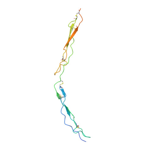
3R6B

Crystal Structure of Thrombospondin-1 TSR Domains 2 and 3


Experimental Data Snapshot

  • Method: X-RAY DIFFRACTION
  • Resolution: 2.40 Å
  • R-Value Free: 0.273 
  • R-Value Work: 0.235 
  • R-Value Observed: 0.239 

wwPDB Validation   3D Report Full Report


This is version 1.3 of the entry. See complete history


Literature

Expression, purification and structural characterization of functionally replete thrombospondin-1 type 1 repeats in a bacterial expression system.

Klenotic, P.A.Page, R.C.Misra, S.Silverstein, R.L.

(2011) Protein Expr Purif 80: 253-259

  • DOI: https://doi.org/10.1016/j.pep.2011.07.010
  • Primary Citation of Related Structures:  
    3R6B

  • PubMed Abstract: 

    The matrix glycoprotein thrombospondin-1 (TSP-1) is a prominent regulator of endothelial cells and angiogenesis. The anti-angiogenic and anti-tumorigenic properties of TSP-1 are in part mediated by the TSP-1 type 1 repeat domains 2 and 3, TSR(2,3). Here, we describe the expression and purification of human TSR(2,3) in milligram quantities from an Escherichia coli expression system. Microvascular endothelial cell migration assays and binding assays with a canonical TSP-1 ligand, histidine-rich glycoprotein (HRGP), indicate that recombinant TSR(2,3) exhibits anti-chemotactic and ligand binding properties similar to full length TSP-1. Furthermore, we determined the structure of E. coli expressed TSR(2,3) by X-ray crystallography at 2.4Å and found the structure to be identical to the existing TSR(2,3) crystal structure determined from a Drosophila expression system. The TSR(2,3) expression and purification protocol developed in this study allows for facile expression of TSR(2,3) for biochemical and biophysical studies, and will aid in the elucidation of the molecular mechanisms of TSP-1 anti-angiogenic and anti-tumorigenic activities.


  • Organizational Affiliation

    Department of Cell Biology, NC10, Lerner Research Institute, Cleveland Clinic, 9500 Euclid Avenue, Cleveland, OH 44195, USA.


Macromolecules
Find similar proteins by:  (by identity cutoff)  |  3D Structure
Entity ID: 1
MoleculeChains Sequence LengthOrganismDetailsImage
Thrombospondin-1153Homo sapiensMutation(s): 0 
Gene Names: THBS1TSPTSP1
UniProt & NIH Common Fund Data Resources
Find proteins for P07996 (Homo sapiens)
Explore P07996 
Go to UniProtKB:  P07996
PHAROS:  P07996
GTEx:  ENSG00000137801 
Entity Groups  
Sequence Clusters30% Identity50% Identity70% Identity90% Identity95% Identity100% Identity
UniProt GroupP07996
Sequence Annotations
Expand
  • Reference Sequence
Experimental Data & Validation

Experimental Data

  • Method: X-RAY DIFFRACTION
  • Resolution: 2.40 Å
  • R-Value Free: 0.273 
  • R-Value Work: 0.235 
  • R-Value Observed: 0.239 
  • Space Group: C 2 2 21
Unit Cell:
Length ( Å )Angle ( ˚ )
a = 66.364α = 90
b = 84.817β = 90
c = 60.707γ = 90
Software Package:
Software NamePurpose
StructureStudiodata collection
PHENIXmodel building
PHENIXrefinement
d*TREKdata reduction
d*TREKdata scaling
PHENIXphasing

Structure Validation

View Full Validation Report


Entry History 

Deposition Data

Revision History  (Full details and data files)

  • Version 1.0: 2011-08-10
    Type: Initial release
  • Version 1.1: 2011-08-17
    Changes: Database references
  • Version 1.2: 2011-11-16
    Changes: Database references
  • Version 1.3: 2023-09-13
    Changes: Data collection, Database references, Derived calculations, Refinement description