3R42 | pdb_00003r42

Crystal structure of the yeast vps23 UEV domain in complex with a vps27 PSDP peptide


Experimental Data Snapshot

  • Method: X-RAY DIFFRACTION
  • Resolution: 1.87 Å
  • R-Value Free: 
    0.203 (Depositor), 0.233 (DCC) 
  • R-Value Work: 
    0.182 (Depositor) 
  • R-Value Observed: 
    0.183 (Depositor) 

Starting Model: experimental
View more details

wwPDB Validation 3D Report Full Report

Validation slider image for 3R42

This is version 1.2 of the entry. See complete history

Literature

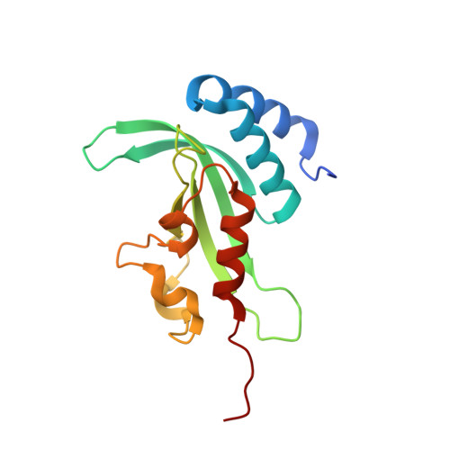
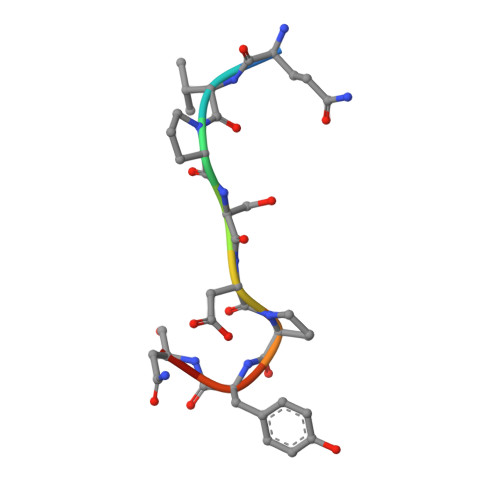
Structural basis for endosomal recruitment of ESCRT-I by ESCRT-0 in yeast.

Ren, X.Hurley, J.H.

(2011) EMBO J 30: 2130-2139

  • DOI: https://doi.org/10.1038/emboj.2011.122
  • Primary Citation Related Structures: 
    3R3Q, 3R42

  • PubMed Abstract: 

    The ESCRT-0 and ESCRT-I complexes coordinate the clustering of ubiquitinated cargo with intralumenal budding of the endosomal membrane, two essential steps in vacuolar/lysosomal protein sorting from yeast to humans. The 1.85-Å crystal structure of interacting regions of the yeast ESCRT-0 and ESCRT-I complexes reveals that PSDP motifs of the Vps27 ESCRT-0 subunit bind to a novel electropositive N-terminal site on the UEV domain of the ESCRT-I subunit Vps23 centred on Trp16. This novel site is completely different from the C-terminal part of the human UEV domain that binds to P(S/T)AP motifs of human ESCRT-0 and HIV-1 Gag. Disruption of the novel PSDP-binding site eliminates the interaction in vitro and blocks enrichment of Vps23 in endosome-related class E compartments in yeast cells. However, this site is non-essential for sorting of the ESCRT cargo Cps1. Taken together, these results show how a conserved motif/domain pair can evolve to use strikingly different binding modes in different organisms.


  • Organizational Affiliation
    • Laboratory of Molecular Biology, National Institute of Diabetes and Digestive and Kidney Diseases, National Institutes of Health, Bethesda, MD, USA.

Macromolecule Content 

  • Total Structure Weight: 19.09 kDa 
  • Atom Count: 1,446 
  • Modeled Residue Count: 164 
  • Deposited Residue Count: 171 
  • Unique protein chains: 2

Macromolecules

Find similar proteins by:|  3D Structure
Entity ID: 1
MoleculeChains  Sequence LengthOrganismDetailsImage
Suppressor protein STP22 of temperature-sensitive alpha-factor receptor and arginine permease162Saccharomyces cerevisiaeMutation(s): 1 
Gene Names: STP22VPS23YCL008CYCL8C
UniProt
Find proteins for P25604 (Saccharomyces cerevisiae (strain ATCC 204508 / S288c))
Explore P25604 
Go to UniProtKB:  P25604
Entity Groups
Sequence Clusters30% Identity50% Identity70% Identity90% Identity95% Identity100% Identity
UniProt GroupP25604
Sequence Annotations
Expand
Reference Sequence
Find similar proteins by:  Sequence   |   3D Structure  
Entity ID: 2
MoleculeChains  Sequence LengthOrganismDetailsImage
Vacuolar protein sorting-associated protein 279Saccharomyces cerevisiaeMutation(s): 0 
UniProt
Find proteins for P40343 (Saccharomyces cerevisiae (strain ATCC 204508 / S288c))
Explore P40343 
Go to UniProtKB:  P40343
Entity Groups
UniProt GroupP40343
Sequence Annotations
Expand
Reference Sequence

Experimental Data & Validation

Experimental Data

  • Method: X-RAY DIFFRACTION
  • Resolution: 1.87 Å
  • R-Value Free:  0.203 (Depositor), 0.233 (DCC) 
  • R-Value Work:  0.182 (Depositor) 
  • R-Value Observed: 0.183 (Depositor) 
Space Group: C 2 2 21
Unit Cell:
Length ( Å )Angle ( ˚ )
a = 95.2α = 90
b = 142.381β = 90
c = 32.065γ = 90
Software Package:
Software NamePurpose
HKL-2000data collection
PHASERphasing
PHENIXrefinement
autoBUSTERrefinement
HKL-2000data reduction
HKL-2000data scaling

Structure Validation

View Full Validation Report


Entry History 

Deposition Data

Revision History  (Full details and data files)

  • Version 1.0: 2011-05-04
    Type: Initial release
  • Version 1.1: 2011-07-13
    Changes: Version format compliance
  • Version 1.2: 2023-09-13
    Changes: Data collection, Database references, Refinement description