Structural basis for the bifunctionality of fructose-1,6-bisphosphate aldolase/phosphatase.
Fushinobu, S., Nishimasu, H., Hattori, D., Song, H.-J., Wakagi, T.(2011) Nature 478: 538-541
- PubMed: 21983966 Search on PubMed
- DOI: https://doi.org/10.1038/nature10457
- Primary Citation Related Structures:
3R1M - PubMed Abstract:
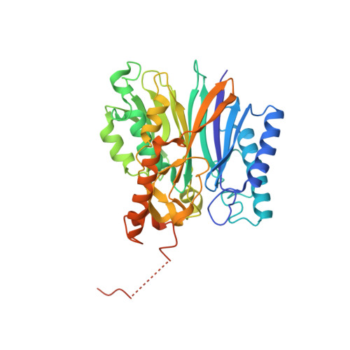
Enzymes catalyse specific reactions and are essential for maintaining life. Although some are referred to as being bifunctional, they consist of either two distinct catalytic domains or a single domain that displays promiscuous substrate specificity. Thus, one enzyme active site is generally responsible for one biochemical reaction. In contrast to this conventional concept, archaeal fructose-1,6-bisphosphate (FBP) aldolase/phosphatase (FBPA/P) consists of a single catalytic domain, but catalyses two chemically distinct reactions of gluconeogenesis: (1) the reversible aldol condensation of dihydroxyacetone phosphate (DHAP) and glyceraldehyde-3-phosphate (GA3P) to FBP; (2) the dephosphorylation of FBP to fructose-6-phosphate (F6P). Thus, FBPA/P is fundamentally different from ordinary enzymes whose active sites are responsible for a specific reaction. However, the molecular mechanism by which FBPA/P achieves its unusual bifunctionality remains unknown. Here we report the crystal structure of FBPA/P at 1.5-Å resolution in the aldolase form, where a critical lysine residue forms a Schiff base with DHAP. A structural comparison of the aldolase form with a previously determined phosphatase form revealed a dramatic conformational change in the active site, demonstrating that FBPA/P metamorphoses its active-site architecture to exhibit dual activities. Thus, our findings expand the conventional concept that one enzyme catalyses one biochemical reaction.
- Department of Biotechnology, Graduate School of Agricultural and Life Sciences, The University of Tokyo, 1-1-1 Yayoi, Bunkyo-ku, Tokyo 113-8657, Japan.
Organizational Affiliation: