3R1D

Crystal structure of GC(8BrG)GCGGCGGC duplex


Experimental Data Snapshot

  • Method: X-RAY DIFFRACTION
  • Resolution: 1.45 Å
  • R-Value Free: 0.270 
  • R-Value Work: 0.232 
  • R-Value Observed: 0.233 

wwPDB Validation   3D Report Full Report


This is version 1.4 of the entry. See complete history


Literature

Crystal structures of CGG RNA repeats with implications for fragile X-associated tremor ataxia syndrome.

Kiliszek, A.Kierzek, R.Krzyzosiak, W.J.Rypniewski, W.

(2011) Nucleic Acids Res 39: 7308-7315

  • DOI: https://doi.org/10.1093/nar/gkr368
  • Primary Citation of Related Structures:  
    3R1C, 3R1D, 3R1E

  • PubMed Abstract: 

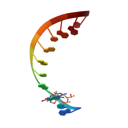
    The CGG repeats are present in the 5'-untranslated region (5'-UTR) of the fragile X mental retardation gene FMR1 and are associated with two diseases: fragile X-associated tremor ataxia syndrome (FXTAS) and fragile X syndrome (FXS). FXTAS occurs when the number of repeats is 55-200 and FXS develops when the number exceeds 200. FXTAS is an RNA-mediated disease in which the expanded CGG tracts form stable structures and sequester important RNA binding proteins. We obtained and analysed three crystal structures of double-helical CGG repeats involving unmodified and 8-Br modified guanosine residues. Despite the presence of the non-canonical base pairs, the helices retain an A-form. In the G-G pairs one guanosine is always in the syn conformation, the other is anti. There are two hydrogen bonds between the Watson-Crick edge of G(anti) and the Hoogsteen edge of G(syn): O6·N1H and N7·N2H. The G(syn)-G(anti) pair shows affinity for binding ions in the major groove. G(syn) causes local unwinding of the helix, compensated elsewhere along the duplex. CGG helical structures appear relatively stable compared with CAG and CUG tracts. This could be an important factor in the RNA's ligand binding affinity and specificity.


  • Organizational Affiliation

    Institute of Bioorganic Chemistry, Polish Academy of Sciences, Noskowskiego 12/14, 61-704 Poznan, Poland.


Macromolecules

Find similar nucleic acids by:  Sequence   |   3D Structure  

Entity ID: 1
MoleculeChains LengthOrganismImage
RNA (5'-R(*GP*CP*(GRB)P*GP*CP*GP*GP*CP*GP*GP*C)-3')
A, B, C, D, E
11N/A
Sequence Annotations
Expand
  • Reference Sequence
Small Molecules
Ligands 2 Unique
IDChains Name / Formula / InChI Key2D Diagram3D Interactions
SO4
Query on SO4

Download Ideal Coordinates CCD File 
G [auth A]
H [auth A]
J [auth B]
K [auth C]
L [auth C]
G [auth A],
H [auth A],
J [auth B],
K [auth C],
L [auth C],
M [auth D],
N [auth E],
O [auth E]
SULFATE ION
O4 S
QAOWNCQODCNURD-UHFFFAOYSA-L
BR
Query on BR

Download Ideal Coordinates CCD File 
F [auth A],
I [auth B]
BROMIDE ION
Br
CPELXLSAUQHCOX-UHFFFAOYSA-M
Experimental Data & Validation

Experimental Data

  • Method: X-RAY DIFFRACTION
  • Resolution: 1.45 Å
  • R-Value Free: 0.270 
  • R-Value Work: 0.232 
  • R-Value Observed: 0.233 
  • Space Group: C 1 2 1
Unit Cell:
Length ( Å )Angle ( ˚ )
a = 118.565α = 90
b = 28.612β = 117.98
c = 61.833γ = 90
Software Package:
Software NamePurpose
MAR345data collection
PHASERphasing
PHENIXrefinement
DENZOdata reduction
SCALEPACKdata scaling

Structure Validation

View Full Validation Report


Entry History 

Deposition Data

Revision History  (Full details and data files)

  • Version 1.0: 2011-06-01
    Type: Initial release
  • Version 1.1: 2011-07-13
    Changes: Version format compliance
  • Version 1.2: 2011-09-14
    Changes: Database references
  • Version 1.3: 2017-11-08
    Changes: Refinement description
  • Version 1.4: 2024-02-21
    Changes: Data collection, Database references, Derived calculations