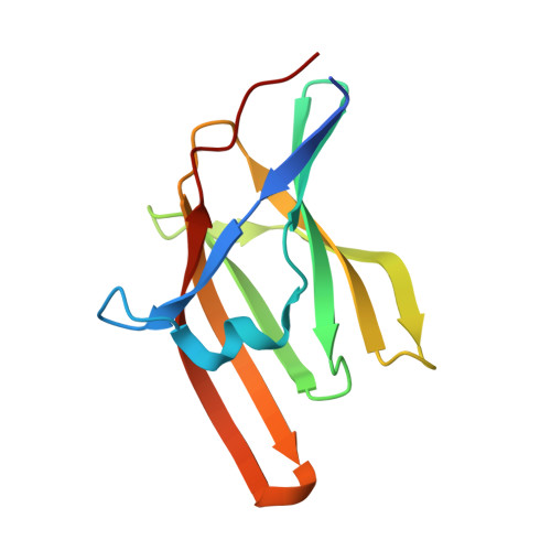
3QZN

Staphylococcus aureus IsdA NEAT domain Y166A variant in complex with heme


Experimental Data Snapshot

  • Method: X-RAY DIFFRACTION
  • Resolution: 2.00 Å
  • R-Value Free: 0.235 
  • R-Value Work: 0.194 
  • R-Value Observed: 0.196 

Starting Model: experimental
View more details

wwPDB Validation   3D Report Full Report


Ligand Structure Quality Assessment 


This is version 1.3 of the entry. See complete history


Literature

Iron-Coordinating Tyrosine Is a Key Determinant of NEAT Domain Heme Transfer.

Grigg, J.C.Mao, C.X.Murphy, M.E.

(2011) J Mol Biol 413: 684-698

  • DOI: https://doi.org/10.1016/j.jmb.2011.08.047
  • Primary Citation of Related Structures:  
    3QZL, 3QZM, 3QZN, 3QZO, 3QZP

  • PubMed Abstract: 

    In humans, heme iron is the most abundant iron source, and bacterial pathogens such as Staphylococcus aureus acquire it for growth. IsdB of S. aureus acquires Fe(III)-protoporphyrin IX (heme) from hemoglobin for transfer to IsdC via IsdA. These three cell-wall-anchored Isd (iron-regulated surface determinant) proteins contain conserved NEAT (near iron transport) domains. The purpose of this work was to delineate the mechanism of heme binding and transfer between the NEAT domains of IsdA, IsdB, and IsdC using a combination of structural and spectroscopic studies. X-ray crystal structures of IsdA NEAT domain (IsdA-N1) variants reveal that removing the native heme-iron ligand Tyr166 is compensated for by iron coordination by His83 on the distal side and that no single mutation of distal loop residues is sufficient to perturb the IsdA-heme complex. Also, alternate heme-iron coordination was observed in structures of IsdA-N1 bound to reduced Fe(II)-protoporphyrin IX and Co(III)-protoporphyrin IX. The IsdA-N1 structural data were correlated with heme transfer kinetics from the NEAT domains of IsdB and IsdC. We demonstrated that the NEAT domains transfer heme at rates comparable to full-length proteins. The second-order rate constant for heme transfer from IsdA-N1 was modestly affected (<2-fold) by the IsdA variants, excluding those at Tyr166. Substituting Tyr166 with Ala or Phe changed the reaction mechanism to one with two observable steps and decreased observed rates >15-fold (to 100-fold excess IsdC). We propose a heme transfer model wherein NEAT domain complexes pass heme iron directly from an iron-coordinating Tyr of the donor protein to the homologous Tyr residues of the acceptor protein.


  • Organizational Affiliation

    Department of Microbiology and Immunology, Life Sciences Institute, The University of British Columbia, Vancouver, BC, Canada V6T 1Z3.


Macromolecules
Find similar proteins by:  (by identity cutoff)  |  3D Structure
Entity ID: 1
MoleculeChains Sequence LengthOrganismDetailsImage
Iron-regulated surface determinant protein A
A, B, C
127Staphylococcus aureus subsp. aureus N315Mutation(s): 1 
Gene Names: frpAisdASA0977stbA
UniProt
Find proteins for Q7A655 (Staphylococcus aureus (strain N315))
Explore Q7A655 
Go to UniProtKB:  Q7A655
Entity Groups  
Sequence Clusters30% Identity50% Identity70% Identity90% Identity95% Identity100% Identity
UniProt GroupQ7A655
Sequence Annotations
Expand
  • Reference Sequence
Small Molecules
Ligands 3 Unique
IDChains Name / Formula / InChI Key2D Diagram3D Interactions
HEM
Query on HEM

Download Ideal Coordinates CCD File 
D [auth A],
G [auth B],
K [auth C]
PROTOPORPHYRIN IX CONTAINING FE
C34 H32 Fe N4 O4
KABFMIBPWCXCRK-RGGAHWMASA-L
SO4
Query on SO4

Download Ideal Coordinates CCD File 
E [auth A],
H [auth B],
I [auth B],
J [auth B]
SULFATE ION
O4 S
QAOWNCQODCNURD-UHFFFAOYSA-L
GOL
Query on GOL

Download Ideal Coordinates CCD File 
F [auth A]GLYCEROL
C3 H8 O3
PEDCQBHIVMGVHV-UHFFFAOYSA-N
Experimental Data & Validation

Experimental Data

  • Method: X-RAY DIFFRACTION
  • Resolution: 2.00 Å
  • R-Value Free: 0.235 
  • R-Value Work: 0.194 
  • R-Value Observed: 0.196 
  • Space Group: P 21 21 21
Unit Cell:
Length ( Å )Angle ( ˚ )
a = 61.302α = 90
b = 96.764β = 90
c = 95.77γ = 90
Software Package:
Software NamePurpose
SCALEPACKdata scaling
MOLREPphasing
REFMACrefinement
PDB_EXTRACTdata extraction
MxDCdata collection
HKL-2000data reduction
HKL-2000data scaling

Structure Validation

View Full Validation Report


Ligand Structure Quality Assessment 


Entry History 

Deposition Data

Revision History  (Full details and data files)

  • Version 1.0: 2011-08-31
    Type: Initial release
  • Version 1.1: 2011-10-05
    Changes: Database references
  • Version 1.2: 2011-11-02
    Changes: Database references
  • Version 1.3: 2023-09-13
    Changes: Data collection, Database references, Derived calculations, Refinement description