3QUJ

Crystal structure of the phosphonate binding protein, PhnD, from Escherichia coli


Experimental Data Snapshot

  • Method: X-RAY DIFFRACTION
  • Resolution: 2.20 Å
  • R-Value Free: 0.276 
  • R-Value Work: 0.202 
  • R-Value Observed: 0.206 

wwPDB Validation   3D Report Full Report


This is version 1.1 of the entry. See complete history


Literature

Structure of the Escherichia coli Phosphonate Binding Protein PhnD and Rationally Optimized Phosphonate Biosensors.

Alicea, I.Marvin, J.S.Miklos, A.E.Ellington, A.D.Looger, L.L.Schreiter, E.R.

(2011) J Mol Biol 414: 356-369

  • DOI: https://doi.org/10.1016/j.jmb.2011.09.047
  • Primary Citation of Related Structures:  
    3P7I, 3QK6, 3QUJ, 3S4U

  • PubMed Abstract: 

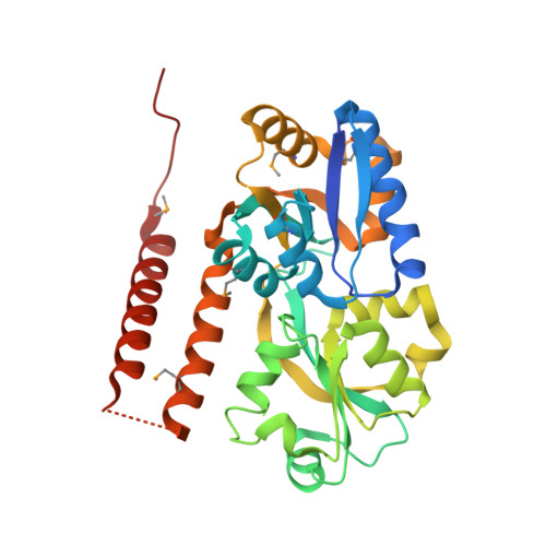
    The phnD gene of Escherichia coli encodes the periplasmic binding protein of the phosphonate (Pn) uptake and utilization pathway. We have crystallized and determined structures of E. coli PhnD (EcPhnD) in the absence of ligand and in complex with the environmentally abundant 2-aminoethylphosphonate (2AEP). Similar to other bacterial periplasmic binding proteins, 2AEP binds near the center of mass of EcPhnD in a cleft formed between two lobes. Comparison of the open, unliganded structure with the closed 2AEP-bound structure shows that the two lobes pivot around a hinge by ~70° between the two states. Extensive hydrogen bonding and electrostatic interactions stabilize 2AEP, which binds to EcPhnD with low nanomolar affinity. These structures provide insight into Pn uptake by bacteria and facilitated the rational design of high signal-to-noise Pn biosensors based on both coupled small-molecule dyes and autocatalytic fluorescent proteins.


  • Organizational Affiliation

    Department of Chemistry, University of Puerto Rico, Río Piedras, San Juan, Puerto Rico 00931.


Macromolecules
Find similar proteins by:  (by identity cutoff)  |  3D Structure
Entity ID: 1
MoleculeChains Sequence LengthOrganismDetailsImage
PhnD, subunit of alkylphosphonate ABC transporter
A, B, C, D
321Escherichia coli UTI89Mutation(s): 0 
Gene Names: phnDUTI89_C4699
UniProt
Find proteins for Q1R3F7 (Escherichia coli (strain UTI89 / UPEC))
Explore Q1R3F7 
Go to UniProtKB:  Q1R3F7
Entity Groups  
Sequence Clusters30% Identity50% Identity70% Identity90% Identity95% Identity100% Identity
UniProt GroupQ1R3F7
Sequence Annotations
Expand
  • Reference Sequence
Small Molecules
Modified Residues  1 Unique
IDChains TypeFormula2D DiagramParent
MSE
Query on MSE
A, B, C, D
L-PEPTIDE LINKINGC5 H11 N O2 SeMET
Experimental Data & Validation

Experimental Data

  • Method: X-RAY DIFFRACTION
  • Resolution: 2.20 Å
  • R-Value Free: 0.276 
  • R-Value Work: 0.202 
  • R-Value Observed: 0.206 
  • Space Group: P 1 21 1
Unit Cell:
Length ( Å )Angle ( ˚ )
a = 79.768α = 90
b = 64.399β = 93.52
c = 107.688γ = 90
Software Package:
Software NamePurpose
SPECdata collection
PHASERphasing
REFMACrefinement
MOSFLMdata reduction
SCALAdata scaling

Structure Validation

View Full Validation Report


Entry History 

Deposition Data

Revision History  (Full details and data files)

  • Version 1.0: 2011-10-12
    Type: Initial release
  • Version 1.1: 2011-12-21
    Changes: Database references