3QU6 | pdb_00003qu6

Crystal structure of IRF-3 DBD free form


Experimental Data Snapshot

  • Method: X-RAY DIFFRACTION
  • Resolution: 2.30 Å
  • R-Value Free: 
    0.263 (Depositor), 0.258 (DCC) 
  • R-Value Work: 
    0.213 (Depositor), 0.211 (DCC) 
  • R-Value Observed: 
    0.216 (Depositor) 

Starting Model: experimental
View more details

wwPDB Validation 3D Report Full Report

Validation slider image for 3QU6

This is version 1.3 of the entry. See complete history

Literature

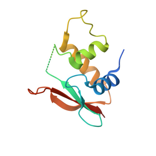
Structures of apo IRF-3 and IRF-7 DNA binding domains: effect of loop L1 on DNA binding.

De Ioannes, P.Escalante, C.R.Aggarwal, A.K.

(2011) Nucleic Acids Res 39: 7300-7307

  • DOI: https://doi.org/10.1093/nar/gkr325
  • Primary Citation Related Structures: 
    3QU3, 3QU6

  • PubMed Abstract: 

    Interferon regulatory factors IRF-3 and IRF-7 are transcription factors essential in the activation of interferon-β (IFN-β) gene in response to viral infections. Although, both proteins recognize the same consensus IRF binding site AANNGAAA, they have distinct DNA binding preferences for sites in vivo. The X-ray structures of IRF-3 and IRF-7 DNA binding domains (DBDs) bound to IFN-β promoter elements revealed flexibility in the loops (L1-L3) and the residues that make contacts with the target sequence. To characterize the conformational changes that occur on DNA binding and how they differ between IRF family members, we have solved the X-ray structures of IRF-3 and IRF-7 DBDs in the absence of DNA. We found that loop L1, carrying the conserved histidine that interacts with the DNA minor groove, is disordered in apo IRF-3 but is ordered in apo IRF-7. This is reflected in differences in DNA binding affinities when the conserved histidine in loop L1 is mutated to alanine in the two proteins. The stability of loop L1 in IRF-7 derives from a unique combination of hydrophobic residues that pack against the protein core. Together, our data show that differences in flexibility of loop L1 are an important determinant of differential IRF-DNA binding.


  • Organizational Affiliation
    • Department of Structural and Chemical Biology, Mount Sinai School of Medicine, Box 1677, 1425 Madison Avenue, NY 10029, USA.

Macromolecule Content 

  • Total Structure Weight: 41.15 kDa 
  • Atom Count: 2,550 
  • Modeled Residue Count: 293 
  • Deposited Residue Count: 348 
  • Unique protein chains: 1

Macromolecules

Find similar proteins by:|  3D Structure
Entity ID: 1
MoleculeChains  Sequence LengthOrganismDetailsImage
IRF3 protein
A, B, C
116Homo sapiensMutation(s): 1 
UniProt & NIH Common Fund Data Resources
Find proteins for Q14653 (Homo sapiens)
Explore Q14653 
Go to UniProtKB:  Q14653
PHAROS:  Q14653
GTEx:  ENSG00000126456 
Entity Groups
Sequence Clusters30% Identity50% Identity70% Identity90% Identity95% Identity100% Identity
UniProt GroupQ14653
Sequence Annotations
Expand
Reference Sequence

Small Molecules

Ligands 3 Unique
IDChains Name / Formula / InChI Key2D Diagram3D Interactions
ZN

Query on ZN


Download:Ideal Coordinates CCD File
D [auth A]
E [auth A]
F [auth A]
G [auth A]
N [auth B]
D [auth A],
E [auth A],
F [auth A],
G [auth A],
N [auth B],
O [auth B],
R [auth C],
S [auth C],
T [auth C]
ZINC ION
Zn
PTFCDOFLOPIGGS-UHFFFAOYSA-N
CL

Query on CL


Download:Ideal Coordinates CCD File
H [auth A]
I [auth A]
J [auth A]
M [auth A]
P [auth B]
H [auth A],
I [auth A],
J [auth A],
M [auth A],
P [auth B],
U [auth C],
V [auth C]
CHLORIDE ION
Cl
VEXZGXHMUGYJMC-UHFFFAOYSA-M
NA

Query on NA


Download:Ideal Coordinates CCD File
K [auth A],
L [auth A],
Q [auth B]
SODIUM ION
Na
FKNQFGJONOIPTF-UHFFFAOYSA-N

Experimental Data & Validation

Experimental Data

  • Method: X-RAY DIFFRACTION
  • Resolution: 2.30 Å
  • R-Value Free:  0.263 (Depositor), 0.258 (DCC) 
  • R-Value Work:  0.213 (Depositor), 0.211 (DCC) 
  • R-Value Observed: 0.216 (Depositor) 
Space Group: P 32 2 1
Unit Cell:
Length ( Å )Angle ( ˚ )
a = 64.908α = 90
b = 64.908β = 90
c = 157.621γ = 120
Software Package:
Software NamePurpose
JDirectordata collection
PHASERphasing
PHENIXrefinement
HKL-2000data reduction
HKL-2000data scaling

Structure Validation

View Full Validation Report


Entry History 

Deposition Data

Revision History  (Full details and data files)

  • Version 1.0: 2011-06-01
    Type: Initial release
  • Version 1.1: 2011-07-13
    Changes: Version format compliance
  • Version 1.2: 2011-09-14
    Changes: Database references
  • Version 1.3: 2023-09-13
    Changes: Data collection, Database references, Derived calculations, Refinement description