3QTL

Structural Basis for Dual-inhibition Mechanism of a Non-classical Kazal-type Serine Protease Inhibitor from Horseshoe Crab in Complex with Subtilisin


Experimental Data Snapshot

  • Method: X-RAY DIFFRACTION
  • Resolution: 2.60 Å
  • R-Value Free: 0.269 
  • R-Value Work: 0.199 
  • R-Value Observed: 0.204 

wwPDB Validation   3D Report Full Report


This is version 1.3 of the entry. See complete history


Literature

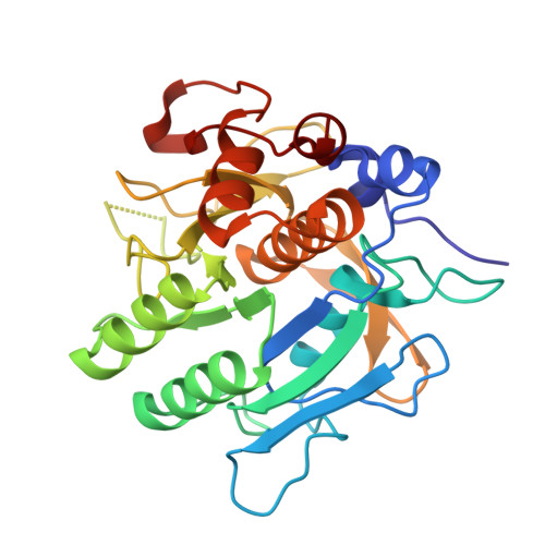
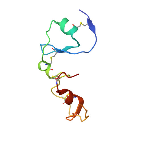
Structural basis for dual-inhibition mechanism of a non-classical kazal-type serine protease inhibitor from horseshoe crab in complex with subtilisin.

Shenoy, R.T.Thangamani, S.Velazquez-Campoy, A.Ho, B.Ding, J.L.Sivaraman, J.

(2011) PLoS One 6: e18838-e18838

  • DOI: https://doi.org/10.1371/journal.pone.0018838
  • Primary Citation of Related Structures:  
    3QTL

  • PubMed Abstract: 

    Serine proteases play a crucial role in host-pathogen interactions. In the innate immune system of invertebrates, multi-domain protease inhibitors are important for the regulation of host-pathogen interactions and antimicrobial activities. Serine protease inhibitors, 9.3-kDa CrSPI isoforms 1 and 2, have been identified from the hepatopancreas of the horseshoe crab, Carcinoscorpius rotundicauda. The CrSPIs were biochemically active, especially CrSPI-1, which potently inhibited subtilisin (Ki = 1.43 nM). CrSPI has been grouped with the non-classical Kazal-type inhibitors due to its unusual cysteine distribution. Here we report the crystal structure of CrSPI-1 in complex with subtilisin at 2.6 Å resolution and the results of biophysical interaction studies. The CrSPI-1 molecule has two domains arranged in an extended conformation. These two domains act as heads that independently interact with two separate subtilisin molecules, resulting in the inhibition of subtilisin activity at a ratio of 1:2 (inhibitor to protease). Each subtilisin molecule interacts with the reactive site loop from each domain of CrSPI-1 through a standard canonical binding mode and forms a single ternary complex. In addition, we propose the substrate preferences of each domain of CrSPI-1. Domain 2 is specific towards the bacterial protease subtilisin, while domain 1 is likely to interact with the host protease, Furin. Elucidation of the structure of the CrSPI-1: subtilisin (1∶2) ternary complex increases our understanding of host-pathogen interactions in the innate immune system at the molecular level and provides new strategies for immunomodulation.


  • Organizational Affiliation

    Department of Biological Sciences, National University of Singapore, Singapore, Singapore.


Macromolecules
Find similar proteins by:  (by identity cutoff)  |  3D Structure
Entity ID: 1
MoleculeChains Sequence LengthOrganismDetailsImage
Subtilisin-like serin protease
A, B, C
274Bacillus licheniformisMutation(s): 0 
Gene Names: aprE
EC: 3.4.21.62
UniProt
Find proteins for Q6PNN5 (Bacillus licheniformis)
Explore Q6PNN5 
Go to UniProtKB:  Q6PNN5
Entity Groups  
Sequence Clusters30% Identity50% Identity70% Identity90% Identity95% Identity100% Identity
UniProt GroupQ6PNN5
Sequence Annotations
Expand
  • Reference Sequence
Find similar proteins by:  (by identity cutoff)  |  3D Structure
Entity ID: 2
MoleculeChains Sequence LengthOrganismDetailsImage
Kazal-type serine protease inhibitor SPI-175Carcinoscorpius rotundicaudaMutation(s): 0 
Gene Names: SPI-1
UniProt
Find proteins for A1X1V8 (Carcinoscorpius rotundicauda)
Explore A1X1V8 
Go to UniProtKB:  A1X1V8
Entity Groups  
Sequence Clusters30% Identity50% Identity70% Identity90% Identity95% Identity100% Identity
UniProt GroupA1X1V8
Sequence Annotations
Expand
  • Reference Sequence
Experimental Data & Validation

Experimental Data

  • Method: X-RAY DIFFRACTION
  • Resolution: 2.60 Å
  • R-Value Free: 0.269 
  • R-Value Work: 0.199 
  • R-Value Observed: 0.204 
  • Space Group: P 1 21 1
Unit Cell:
Length ( Å )Angle ( ˚ )
a = 73.84α = 90
b = 65.072β = 95.44
c = 111.909γ = 90
Software Package:
Software NamePurpose
DENZOdata reduction
SCALEPACKdata scaling
MOLREPphasing
REFMACrefinement
PDB_EXTRACTdata extraction
CBASSdata collection
PHENIXrefinement

Structure Validation

View Full Validation Report


Entry History 

Deposition Data

Revision History  (Full details and data files)

  • Version 1.0: 2011-06-01
    Type: Initial release
  • Version 1.1: 2011-07-13
    Changes: Version format compliance
  • Version 1.2: 2017-11-08
    Changes: Refinement description
  • Version 1.3: 2023-11-01
    Changes: Data collection, Database references, Refinement description