A strategy for antagonizing quorum sensing.
Chen, G., Swem, L.R., Swem, D.L., Stauff, D.L., O'Loughlin, C.T., Jeffrey, P.D., Bassler, B.L., Hughson, F.M.(2011) Mol Cell 42: 199-209
- PubMed: 21504831 Search on PubMedSearch on PubMed Central
- DOI: https://doi.org/10.1016/j.molcel.2011.04.003
- Primary Citation Related Structures:
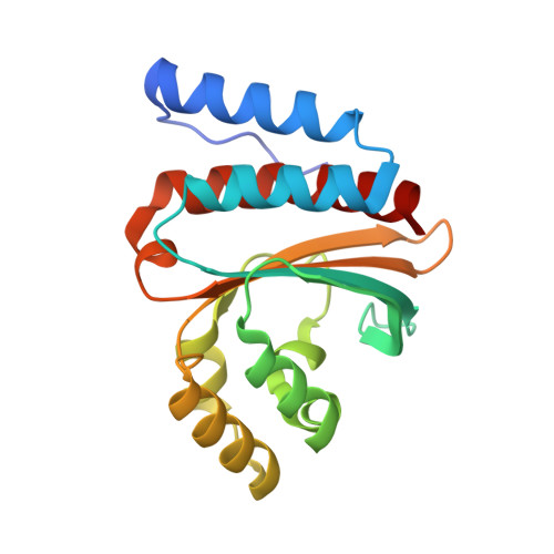
3QP1, 3QP2, 3QP4, 3QP5, 3QP6, 3QP8 - PubMed Abstract:
Quorum-sensing bacteria communicate via small molecules called autoinducers to coordinate collective behaviors. Because quorum sensing controls virulence factor expression in many clinically relevant pathogens, membrane-permeable quorum sensing antagonists that prevent population-wide expression of virulence genes offer a potential route to novel antibacterial therapeutics. Here, we report a strategy for inhibiting quorum-sensing receptors of the widespread LuxR family. Structure-function studies with natural and synthetic ligands demonstrate that the dimeric LuxR-type transcription factor CviR from Chromobacterium violaceum is potently antagonized by molecules that bind in place of the native acylated homoserine lactone autoinducer, provided that they stabilize a closed conformation. In such conformations, each of the two DNA-binding domains interacts with the ligand-binding domain of the opposing monomer. Consequently, the DNA-binding helices are held apart by ∼60 Å, twice the ∼30 Å separation required for operator binding. This approach may represent a general strategy for the inhibition of multidomain proteins.
- Department of Molecular Biology, Princeton University, Princeton, NJ 08544, USA.
Organizational Affiliation: