3QOL

Crystal structure of Staphylococcal nuclease variant D+PHS/V23E at pH 6 determined at 100 K


Experimental Data Snapshot

  • Method: X-RAY DIFFRACTION
  • Resolution: 1.90 Å
  • R-Value Free: 0.227 
  • R-Value Work: 0.187 
  • R-Value Observed: 0.189 

wwPDB Validation   3D Report Full Report


Ligand Structure Quality Assessment 


This is version 1.4 of the entry. See complete history


Literature

Structural and thermodynamic consequences of burial of an artificial ion pair in the hydrophobic interior of a protein.

Robinson, A.C.Castaneda, C.A.Schlessman, J.L.Garcia-Moreno E, B.

(2014) Proc Natl Acad Sci U S A 111: 11685-11690

  • DOI: https://doi.org/10.1073/pnas.1402900111
  • Primary Citation of Related Structures:  
    3NHH, 3QOL

  • PubMed Abstract: 

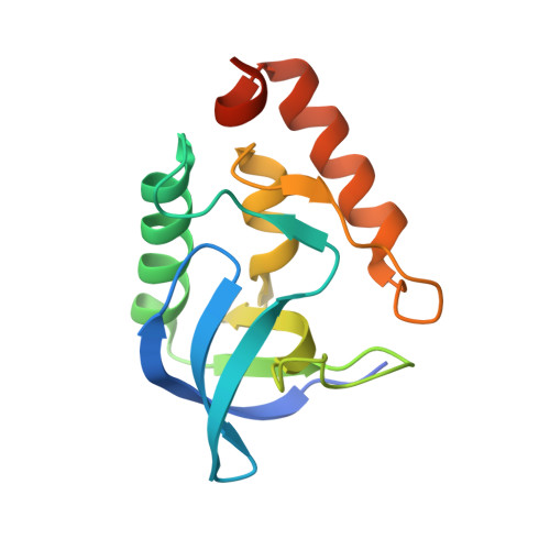
    An artificial charge pair buried in the hydrophobic core of staphylococcal nuclease was engineered by making the V23E and L36K substitutions. Buried individually, Glu-23 and Lys-36 both titrate with pKa values near 7. When buried together their pKa values appear to be normal. The ionizable moieties of the buried Glu-Lys pair are 2.6 Å apart. The interaction between them at pH 7 is worth 5 kcal/mol. Despite this strong interaction, the buried Glu-Lys pair destabilizes the protein significantly because the apparent Coulomb interaction is sufficient to offset the dehydration of only one of the two buried charges. Save for minor reorganization of dipoles and water penetration consistent with the relatively high dielectric constant reported by the buried ion pair, there is no evidence that the presence of two charges in the hydrophobic interior of the protein induces any significant structural reorganization. The successful engineering of an artificial ion pair in a highly hydrophobic environment suggests that buried Glu-Lys pairs in dehydrated environments can be charged and that it is possible to engineer charge clusters that loosely resemble catalytic sites in a scaffold protein with high thermodynamic stability, without the need for specialized structural adaptations.


  • Organizational Affiliation

    Department of Biophysics, Johns Hopkins University, Baltimore, MD 21218; and.


Macromolecules
Find similar proteins by:  (by identity cutoff)  |  3D Structure
Entity ID: 1
MoleculeChains Sequence LengthOrganismDetailsImage
Thermonuclease143Staphylococcus aureusMutation(s): 6 
Gene Names: nuc
EC: 3.1.31.1
UniProt
Find proteins for Q8NXI6 (Staphylococcus aureus (strain MW2))
Explore Q8NXI6 
Go to UniProtKB:  Q8NXI6
Entity Groups  
Sequence Clusters30% Identity50% Identity70% Identity90% Identity95% Identity100% Identity
UniProt GroupQ8NXI6
Sequence Annotations
Expand
  • Reference Sequence
Small Molecules
Ligands 2 Unique
IDChains Name / Formula / InChI Key2D Diagram3D Interactions
THP
Query on THP

Download Ideal Coordinates CCD File 
C [auth A]THYMIDINE-3',5'-DIPHOSPHATE
C10 H16 N2 O11 P2
CSNCBOPUCJOHLS-XLPZGREQSA-N
CA
Query on CA

Download Ideal Coordinates CCD File 
B [auth A]CALCIUM ION
Ca
BHPQYMZQTOCNFJ-UHFFFAOYSA-N
Experimental Data & Validation

Experimental Data

  • Method: X-RAY DIFFRACTION
  • Resolution: 1.90 Å
  • R-Value Free: 0.227 
  • R-Value Work: 0.187 
  • R-Value Observed: 0.189 
  • Space Group: P 1 21 1
Unit Cell:
Length ( Å )Angle ( ˚ )
a = 31.033α = 90
b = 60.453β = 94.44
c = 37.626γ = 90
Software Package:
Software NamePurpose
APEXdata collection
PHASERphasing
REFMACrefinement
SAINTdata reduction
XPREPdata reduction

Structure Validation

View Full Validation Report


Ligand Structure Quality Assessment 


Entry History 

Deposition Data

Revision History  (Full details and data files)

  • Version 1.0: 2011-03-16
    Type: Initial release
  • Version 1.1: 2011-07-13
    Changes: Version format compliance
  • Version 1.2: 2014-08-20
    Changes: Database references
  • Version 1.3: 2014-09-03
    Changes: Database references
  • Version 1.4: 2023-09-13
    Changes: Data collection, Database references, Derived calculations, Refinement description