3QMG | pdb_00003qmg

Structural Basis of Selective Binding of Non-Methylated CpG islands by the CXXC Domain of CFP1


Experimental Data Snapshot

  • Method: X-RAY DIFFRACTION
  • Resolution: 2.30 Å
  • R-Value Free: 
    0.279 (Depositor), 0.266 (DCC) 
  • R-Value Work: 
    0.216 (Depositor), 0.216 (DCC) 
  • R-Value Observed: 
    0.219 (Depositor) 

Starting Model: experimental
View more details

wwPDB Validation 3D Report Full Report

Validation slider image for 3QMG

This is version 1.2 of the entry. See complete history

Literature

The structural basis for selective binding of non-methylated CpG islands by the CFP1 CXXC domain.

Xu, C.Bian, C.Lam, R.Dong, A.Min, J.

(2011) Nat Commun 2: 227-227

  • DOI: https://doi.org/10.1038/ncomms1237
  • Primary Citation Related Structures: 
    3QMB, 3QMC, 3QMD, 3QMG, 3QMH, 3QMI

  • PubMed Abstract: 

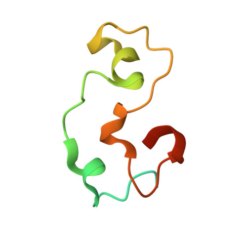
    CFP1 is a CXXC domain-containing protein and an essential component of the SETD1 histone H3K4 methyltransferase complex. CXXC domain proteins direct different chromatin-modifying activities to various chromatin regions. Here, we report crystal structures of the CFP1 CXXC domain in complex with six different CpG DNA sequences. The crescent-shaped CFP1 CXXC domain is wedged into the major groove of the CpG DNA, distorting the B-form DNA, and interacts extensively with the major groove of the DNA. The structures elucidate the molecular mechanism of the non-methylated CpG-binding specificity of the CFP1 CXXC domain. The CpG motif is confined by a tripeptide located in a rigid loop, which only allows the accommodation of the non-methylated CpG dinucleotide. Furthermore, we demonstrate that CFP1 has a preference for a guanosine nucleotide following the CpG motif.


  • Organizational Affiliation
    • Structural Genomics Consortium, University of Toronto, Toronto, Ontario, Canada.

Macromolecule Content 

  • Total Structure Weight: 17.09 kDa 
  • Atom Count: 911 
  • Modeled Residue Count: 76 
  • Deposited Residue Count: 103 
  • Unique protein chains: 1
  • Unique nucleic acid chains: 2

Macromolecules


Find similar proteins by:|  3D Structure
Entity ID: 1
MoleculeChains  Sequence LengthOrganismDetailsImage
CpG-binding protein79Homo sapiensMutation(s): 0 
Gene Names: CFP1CGBPCXXC1PCCX1PHF18
UniProt & NIH Common Fund Data Resources
Find proteins for Q9P0U4 (Homo sapiens)
Explore Q9P0U4 
Go to UniProtKB:  Q9P0U4
PHAROS:  Q9P0U4
GTEx:  ENSG00000154832 
Entity Groups
Sequence Clusters30% Identity50% Identity70% Identity90% Identity95% Identity100% Identity
UniProt GroupQ9P0U4
Sequence Annotations
Expand
Reference Sequence
Find similar nucleic acids by:  Sequence
Entity ID: 2
MoleculeChains LengthOrganismImage
5'-D(*GP*CP*CP*AP*AP*CP*GP*GP*TP*GP*GP*C)-3'12N/A
Sequence Annotations
Expand
Reference Sequence
Find similar nucleic acids by:  Sequence
Entity ID: 3
MoleculeChains LengthOrganismImage
5'-D(*GP*CP*CP*AP*CP*CP*GP*TP*TP*GP*GP*C)-3'12N/A
Sequence Annotations
Expand
Reference Sequence

Experimental Data & Validation

Experimental Data

  • Method: X-RAY DIFFRACTION
  • Resolution: 2.30 Å
  • R-Value Free:  0.279 (Depositor), 0.266 (DCC) 
  • R-Value Work:  0.216 (Depositor), 0.216 (DCC) 
  • R-Value Observed: 0.219 (Depositor) 
Space Group: C 2 2 21
Unit Cell:
Length ( Å )Angle ( ˚ )
a = 30.436α = 90
b = 74.91β = 90
c = 125.755γ = 90
Software Package:
Software NamePurpose
REFMACrefinement

Structure Validation

View Full Validation Report


Entry History 

Deposition Data

Revision History  (Full details and data files)

  • Version 1.0: 2011-02-23
    Type: Initial release
  • Version 1.1: 2011-07-13
    Changes: Version format compliance
  • Version 1.2: 2023-09-13
    Changes: Data collection, Database references, Derived calculations, Refinement description