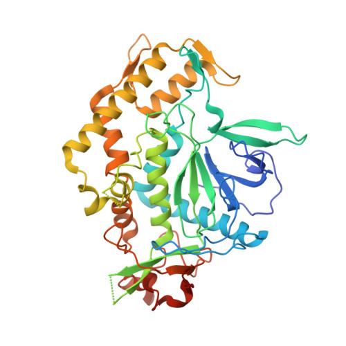
3QJ0

Crystal Structure of BoNT/A LC complexed with Hydroxamate-based Inhibitor PT-3


Experimental Data Snapshot

  • Method: X-RAY DIFFRACTION
  • Resolution: 2.30 Å
  • R-Value Free: 0.229 
  • R-Value Work: 0.198 
  • R-Value Observed: 0.200 

wwPDB Validation   3D Report Full Report


Ligand Structure Quality Assessment 


This is version 1.3 of the entry. See complete history


Literature

Structural Characterization of Three Novel Hydroxamate-Based Zinc Chelating Inhibitors of the Clostridium botulinum Serotype A Neurotoxin Light Chain Metalloprotease Reveals a Compact Binding Site Resulting from 60/70 Loop Flexibility.

Thompson, A.A.Jiao, G.S.Kim, S.Thai, A.Cregar-Hernandez, L.Margosiak, S.A.Johnson, A.T.Han, G.W.O'Malley, S.Stevens, R.C.

(2011) Biochemistry 50: 4019-4028

  • DOI: https://doi.org/10.1021/bi2001483
  • Primary Citation of Related Structures:  
    3QIX, 3QIY, 3QIZ, 3QJ0

  • PubMed Abstract: 

    Neurotoxins synthesized by Clostridium botulinum bacteria (BoNT), the etiological agent of human botulism, are extremely toxic proteins making them high-risk agents for bioterrorism. Small molecule inhibitor development has been focused on the light chain zinc-dependent metalloprotease domain of the neurotoxin, an effort that has been hampered by its relatively flexible active site. Developed in concert with structure--activity relationship studies, the X-ray crystal structures of the complex of BoNT serotype A light chain (BoNT/A LC) with three different micromolar-potency hydroxamate-based inhibitors are reported here. Comparison with an unliganded BoNT/A LC structure reveals significant changes in the active site as a result of binding by the unique inhibitor scaffolds. The 60/70 loop at the opening of the active site pocket undergoes the largest conformational change, presumably through an induced-fit mechanism, resulting in the most compact catalytic pocket observed in all known BoNT/A LC structures.


  • Organizational Affiliation

    Department of Molecular Biology, The Scripps Research Institute, La Jolla, California 92037, United States.


Macromolecules
Find similar proteins by:  (by identity cutoff)  |  3D Structure
Entity ID: 1
MoleculeChains Sequence LengthOrganismDetailsImage
Botulinum neurotoxin type A430Clostridium botulinum A str. HallMutation(s): 0 
Gene Names: botACBO0806CLC_0862Neurotoxin Light Chain
EC: 3.4.24.69
Entity Groups  
Sequence Clusters30% Identity50% Identity70% Identity90% Identity95% Identity100% Identity
Sequence Annotations
Expand
  • Reference Sequence
Small Molecules
Binding Affinity Annotations 
IDSourceBinding Affinity
QI3 Binding MOAD:  3QJ0 Ki: 6300 (nM) from 1 assay(s)
PDBBind:  3QJ0 Ki: 6300 (nM) from 1 assay(s)
Experimental Data & Validation

Experimental Data

  • Method: X-RAY DIFFRACTION
  • Resolution: 2.30 Å
  • R-Value Free: 0.229 
  • R-Value Work: 0.198 
  • R-Value Observed: 0.200 
  • Space Group: P 21 21 2
Unit Cell:
Length ( Å )Angle ( ˚ )
a = 59.17α = 90
b = 188.918β = 90
c = 42.562γ = 90
Software Package:
Software NamePurpose
DENZOdata reduction
SCALEPACKdata scaling
PHASERphasing
PHENIXrefinement
PDB_EXTRACTdata extraction
Blu-Icedata collection
HKL-2000data reduction
HKL-2000data scaling

Structure Validation

View Full Validation Report


Ligand Structure Quality Assessment 


Entry History 

Deposition Data

Revision History  (Full details and data files)

  • Version 1.0: 2011-04-13
    Type: Initial release
  • Version 1.1: 2011-07-13
    Changes: Version format compliance
  • Version 1.2: 2017-11-08
    Changes: Refinement description
  • Version 1.3: 2023-09-13
    Changes: Data collection, Database references, Derived calculations, Refinement description