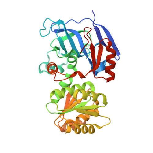
3QE3

Sheep liver sorbitol dehydrogenase


Experimental Data Snapshot

  • Method: X-RAY DIFFRACTION
  • Resolution: 1.90 Å
  • R-Value Free: 0.234 
  • R-Value Work: 0.192 
  • R-Value Observed: 0.195 

wwPDB Validation   3D Report Full Report


This is version 1.2 of the entry. See complete history


Literature

X-ray crystal structure and small-angle X-ray scattering of sheep liver sorbitol dehydrogenase.

Yennawar, H.Moller, M.Gillilan, R.Yennawar, N.

(2011) Acta Crystallogr D Biol Crystallogr 67: 440-446

  • DOI: 10.1107/S0907444911007815
  • Primary Citation of Related Structures:  
    3QE3

  • PubMed Abstract: 
  • The X-ray crystal structure of sheep liver sorbitol dehydrogenase (slSDH) has been determined using the crystal structure of human sorbitol dehydrogenase (hSDH) as a molecular-replacement model. slSDH crystallized in space group I222 with one monomer in the asymmetric unit ...

    The X-ray crystal structure of sheep liver sorbitol dehydrogenase (slSDH) has been determined using the crystal structure of human sorbitol dehydrogenase (hSDH) as a molecular-replacement model. slSDH crystallized in space group I222 with one monomer in the asymmetric unit. A conserved tetramer that superposes well with that seen in hSDH (despite belonging to a different space group) and obeying the 222 crystal symmetry is seen in slSDH. An acetate molecule is bound in the active site, coordinating to the active-site zinc through a water molecule. Glycerol, a substrate of slSDH, also occupies the substrate-binding pocket together with the acetate designed by nature to fit large polyol substrates. The substrate-binding pocket is seen to be in close proximity to the tetramer interface, which explains the need for the structural integrity of the tetramer for enzyme activity. Small-angle X-ray scattering was also used to identify the quaternary structure of the tetramer of slSDH in solution.


    Organizational Affiliation

    Department of Biochemistry and Molecular Biology and Huck Institutes of Life Sciences, Pennsylvania State University, 8 Althouse Laboratory, University Park, PA 16802, USA.


Macromolecules
Find similar proteins by:  (by identity cutoff)  |  3D Structure
Entity ID: 1
MoleculeChainsSequence LengthOrganismDetailsImage
Sorbitol dehydrogenaseA355Ovis ariesMutation(s): 0 
Gene Names: SORD
EC: 1.1.1.14 (PDB Primary Data), 1.1.1 (UniProt), 1.1.1.9 (UniProt)
UniProt
Find proteins for P07846 (Ovis aries)
Explore P07846 
Go to UniProtKB:  P07846
Entity Groups  
Sequence Clusters30% Identity50% Identity70% Identity90% Identity95% Identity100% Identity
UniProt GroupP07846
Protein Feature View
Expand
  • Reference Sequence
Small Molecules
Ligands 3 Unique
IDChainsName / Formula / InChI Key2D Diagram3D Interactions
GOL
Query on GOL

Download Ideal Coordinates CCD File 
C [auth A]GLYCEROL
C3 H8 O3
PEDCQBHIVMGVHV-UHFFFAOYSA-N
 Ligand Interaction
ZN
Query on ZN

Download Ideal Coordinates CCD File 
B [auth A]ZINC ION
Zn
PTFCDOFLOPIGGS-UHFFFAOYSA-N
 Ligand Interaction
ACY
Query on ACY

Download Ideal Coordinates CCD File 
D [auth A],
E [auth A],
F [auth A]
ACETIC ACID
C2 H4 O2
QTBSBXVTEAMEQO-UHFFFAOYSA-N
 Ligand Interaction
Experimental Data & Validation

Experimental Data

  • Method: X-RAY DIFFRACTION
  • Resolution: 1.90 Å
  • R-Value Free: 0.234 
  • R-Value Work: 0.192 
  • R-Value Observed: 0.195 
  • Space Group: I 2 2 2
Unit Cell:
Length ( Å )Angle ( ˚ )
a = 67.749α = 90
b = 85.881β = 90
c = 119.888γ = 90
Software Package:
Software NamePurpose
CrystalCleardata collection
PHENIXmodel building
PHENIXrefinement
HKL-2000data reduction
HKL-2000data scaling
PHENIXphasing

Structure Validation

View Full Validation Report


Entry History 

Deposition Data

Revision History  (Full details and data files)

  • Version 1.0: 2011-04-27
    Type: Initial release
  • Version 1.1: 2011-07-13
    Changes: Version format compliance
  • Version 1.2: 2013-03-27
    Changes: Database references