3QCN | pdb_00003qcn

Human receptor protein tyrosine phosphatase gamma, domain 1, trigonal crystal form


Experimental Data Snapshot

  • Method: X-RAY DIFFRACTION
  • Resolution: 2.41 Å
  • R-Value Free: 
    0.234 (Depositor), 0.240 (DCC) 
  • R-Value Work: 
    0.198 (Depositor), 0.200 (DCC) 
  • R-Value Observed: 
    0.199 (Depositor) 

Starting Model: experimental
View more details

wwPDB Validation 3D Report Full Report

Validation slider image for 3QCN

This is version 1.1 of the entry. See complete history

Literature

Small molecule receptor protein tyrosine phosphatase [gamma](RPTP[gamma]) ligands that inhibit phosphatase activity via perturbation of the tryptophan-proline-aspartate (WPD) loop

Sheriff, S.Beno, B.R.Zhai, W.Kostich, W.A.McDonnell, P.A.Kish, K.Goldfarb, V.Gao, M.Kiefer, S.E.Yanchunas, J.Huang, Y.Shi, S.Zhu, S.Dzierba, C.Bronson, J.Macor, J.E.Appiah, K.K.Westphal, R.S.O'Connell, J.Gerritz, S.W.

(2011) J Med Chem 54: 6548-6562

  • DOI: https://doi.org/10.1021/jm2003766
  • Primary Citation Related Structures: 
    3QCB, 3QCC, 3QCD, 3QCE, 3QCF, 3QCG, 3QCH, 3QCI, 3QCJ, 3QCK, 3QCL, 3QCM, 3QCN

  • PubMed Abstract: 

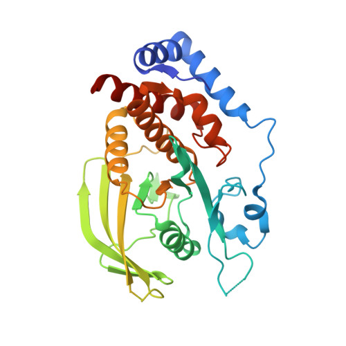
    Protein tyrosine phosphatases (PTPs) catalyze the dephosphorylation of tyrosine residues, a process that involves a conserved tryptophan-proline-aspartate (WPD) loop in catalysis. In previously determined structures of PTPs, the WPD-loop has been observed in either an "open" conformation or a "closed" conformation. In the current work, X-ray structures of the catalytic domain of receptor-like protein tyrosine phosphatase γ (RPTPγ) revealed a ligand-induced "superopen" conformation not previously reported for PTPs. In the superopen conformation, the ligand acts as an apparent competitive inhibitor and binds in a small hydrophobic pocket adjacent to, but distinct from, the active site. In the open and closed WPD-loop conformations of RPTPγ, the side chain of Trp1026 partially occupies this pocket. In the superopen conformation, Trp1026 is displaced allowing a 3,4-dichlorobenzyl substituent to occupy this site. The bound ligand prevents closure of the WPD-loop over the active site and disrupts the catalytic cycle of the enzyme.


  • Organizational Affiliation
    • Bristol-Myers Squibb Research and Development, P.O. Box 4000, Princeton, New Jersey 08543-4000, United States. steven.sheriff@bms.com

Macromolecule Content 

  • Total Structure Weight: 35.71 kDa 
  • Atom Count: 2,409 
  • Modeled Residue Count: 286 
  • Deposited Residue Count: 310 
  • Unique protein chains: 1

Macromolecules

Find similar proteins by:|  3D Structure
Entity ID: 1
MoleculeChains  Sequence LengthOrganismDetailsImage
Receptor-type tyrosine-protein phosphatase gamma310Homo sapiensMutation(s): 2 
Gene Names: PTPGPTPRG
EC: 3.1.3.48
UniProt & NIH Common Fund Data Resources
Find proteins for P23470 (Homo sapiens)
Explore P23470 
Go to UniProtKB:  P23470
PHAROS:  P23470
GTEx:  ENSG00000144724 
Entity Groups
Sequence Clusters30% Identity50% Identity70% Identity90% Identity95% Identity100% Identity
UniProt GroupP23470
Sequence Annotations
Expand
Reference Sequence

Experimental Data & Validation

Experimental Data

  • Method: X-RAY DIFFRACTION
  • Resolution: 2.41 Å
  • R-Value Free:  0.234 (Depositor), 0.240 (DCC) 
  • R-Value Work:  0.198 (Depositor), 0.200 (DCC) 
  • R-Value Observed: 0.199 (Depositor) 
Space Group: P 32 2 1
Unit Cell:
Length ( Å )Angle ( ˚ )
a = 75.2α = 90
b = 75.2β = 90
c = 152.3γ = 120
Software Package:
Software NamePurpose
AMoREphasing
BUSTERrefinement
HKL-2000data reduction
HKL-2000data scaling

Structure Validation

View Full Validation Report


Entry History 

Deposition Data

  • Released Date: 2011-12-28 
  • Deposition Author(s): Sheriff, S.

Revision History  (Full details and data files)

  • Version 1.0: 2011-12-28
    Type: Initial release
  • Version 1.1: 2023-09-13
    Changes: Data collection, Database references, Refinement description