


Crystal structure of phosphoserine aminotransferase from Yersinia pestis CO92
Nocek, B., Maltseva, N., Papazisi, L., Anderson, W., Joachimiak, A.To be published.
Experimental Data Snapshot
Entity ID: 1 | |||||
|---|---|---|---|---|---|
| Molecule | Chains | Sequence Length | Organism | Details | Image |
| Phosphoserine aminotransferase | 364 | Yersinia pestis CO92 | Mutation(s): 0 EC: 2.6.1.52 | 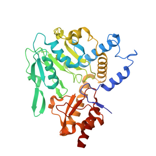 | |
| Ligands 1 Unique | |||||
|---|---|---|---|---|---|
| ID | Chains | Name / Formula / InChI Key | 2D Diagram | 3D Interactions | |
| PLP Download:Ideal Coordinates CCD File | C [auth A], D [auth B] | PYRIDOXAL-5'-PHOSPHATE C8 H10 N O6 P NGVDGCNFYWLIFO-UHFFFAOYSA-N |  | ||
| Length ( Å ) | Angle ( ˚ ) |
|---|---|
| a = 78.338 | α = 90 |
| b = 87.951 | β = 90 |
| c = 106.177 | γ = 90 |
| Software Name | Purpose |
|---|---|
| SBC-Collect | data collection |
| MOLREP | phasing |
| REFMAC | refinement |
| HKL-3000 | data reduction |
| HKL-3000 | data scaling |


