


Crystal structure of dipeptidyl peptidase IV in complex with inhibitor
Liu, S.P.To be published.
Experimental Data Snapshot
Entity ID: 1 | |||||
|---|---|---|---|---|---|
| Molecule | Chains | Sequence Length | Organism | Details | Image |
| Dipeptidyl peptidase 4 | 748 | Homo sapiens | Mutation(s): 2 Gene Names: DPP4, ADCP2, CD26 EC: 3.4.14.5 | 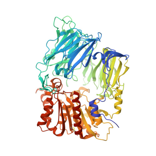 | |
UniProt & NIH Common Fund Data Resources | |||||
PHAROS: P27487 GTEx: ENSG00000197635 | |||||
Entity Groups | |||||
| Sequence Clusters | 30% Identity50% Identity70% Identity90% Identity95% Identity100% Identity | ||||
| UniProt Group | P27487 | ||||
Glycosylation | |||||
| Glycosylation Sites: 2 | Go to GlyGen: P27487-1 | ||||
Sequence AnnotationsExpand | |||||
Reference Sequence | |||||
Entity ID: 2 | |||||
|---|---|---|---|---|---|
| Molecule | Chains | Length | 2D Diagram | Glycosylation | D Interactions |
| 2-acetamido-2-deoxy-beta-D-glucopyranose-(1-4)-2-acetamido-2-deoxy-beta-D-glucopyranose | C, E | 2 |  | N-Glycosylation | |
Glycosylation Resources | |||||
GlyTouCan: G42666HT GlyCosmos: G42666HT GlyGen: G42666HT | |||||
Entity ID: 3 | |||||
|---|---|---|---|---|---|
| Molecule | Chains | Length | 2D Diagram | Glycosylation | D Interactions |
| alpha-D-mannopyranose-(1-3)-beta-D-mannopyranose-(1-4)-2-acetamido-2-deoxy-beta-D-glucopyranose-(1-4)-2-acetamido-2-deoxy-beta-D-glucopyranose | D | 4 |  | N-Glycosylation | |
Glycosylation Resources | |||||
GlyTouCan: G81315DD GlyCosmos: G81315DD GlyGen: G81315DD | |||||
| Ligands 2 Unique | |||||
|---|---|---|---|---|---|
| ID | Chains | Name / Formula / InChI Key | 2D Diagram | 3D Interactions | |
| NXZ Download:Ideal Coordinates CCD File | H [auth A], J [auth B] | 1-[(3S,4S)-4-amino-1-(6-phenylpyrimidin-4-yl)pyrrolidin-3-yl]piperidin-2-one C19 H23 N5 O FHAABBQZMYZFKY-RDJZCZTQSA-N |  | ||
| NAG Download:Ideal Coordinates CCD File | F [auth A], G [auth A], I [auth B] | 2-acetamido-2-deoxy-beta-D-glucopyranose C8 H15 N O6 OVRNDRQMDRJTHS-FMDGEEDCSA-N |  | ||
| Length ( Å ) | Angle ( ˚ ) |
|---|---|
| a = 65.751 | α = 90 |
| b = 69.082 | β = 90 |
| c = 424.16 | γ = 90 |
| Software Name | Purpose |
|---|---|
| SCALA | data scaling |
| BUSTER-TNT | refinement |
| PDB_EXTRACT | data extraction |
| XSCALE | data scaling |
| BUSTER | refinement |


