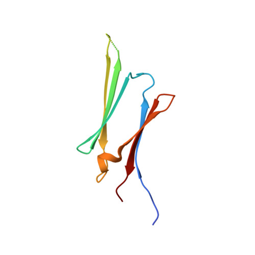
Three-Dimensional Structure of alpha-Crystallin Domain Dimers of Human Small Heat Shock Proteins HSPB1 and HSPB6
Baranova, E.V., Weeks, S.D., Beelen, S., Bukach, O.V., Gusev, N.B., Strelkov, S.V.(2011) J Mol Biology
- PubMed: 21641913 Search on PubMed
- DOI: https://doi.org/10.1016/j.jmb.2011.05.024
- Primary Citation Related Structures:
3Q9P, 3Q9Q - PubMed Abstract:
Small heat shock proteins (sHSPs) are a family of evolutionary conserved ATP-independent chaperones. These proteins share a common architecture defined by a signature α-crystallin domain (ACD) flanked by highly variable N- and C-terminal extensions. The ACD, which has an immunoglobulin-like fold, plays an important role in sHSP assembly. This domain mediates dimer formation of individual protomers, which then may assemble into larger oligomers. In vertebrate sHSPs, the dimer interface is formed by the symmetrical antiparallel pairing of two β-strands (β7), generating an extended β-sheet on one face of the ACD dimer. Recent structural studies of isolated ACDs from a number of vertebrate sHSPs suggest a variability in the register of the β7/β7 strand interface, which may, in part, give rise to the polydispersity often associated with the full-length proteins. To further analyze the structure of ACD dimers, we have employed a combination of X-ray crystallography and solution small-angle X-ray scattering (SAXS) to study the ACD-containing fragments of human HSPB1 (HSP27) and HSPB6 (HSP20). Unexpectedly, the obtained crystal structure of the HSPB1 fragment does not reveal the typical β7/β7 dimers but, rather, hexamers formed by an asymmetric contact between the β4 and the β7 strands from adjacent ACDs. Nevertheless, in solution, both ACDs form stable dimers via the symmetric antiparallel interaction of β7 strands. Using SAXS, we show that it is possible to discriminate between different putative registers of the β7/β7 interface, with the results indicating that, under physiological conditions, there is only a single register of the strands for both proteins.
- Laboratory for Biocrystallography, Department of Pharmaceutical Sciences, Katholieke Universiteit Leuven, 3000 Leuven, Belgium.
Organizational Affiliation: