


An Uncommon Crystal Structure of a Marine Knottin Peptide from Asteropus sp.
Li, H., Bowling, J.J., Fronczek, F.R., Hong, J., Hamann, M.T., Jung, J.H.To be published.
Experimental Data Snapshot
wwPDB Validation 3D Report Full Report

Entity ID: 1 | |||||
|---|---|---|---|---|---|
| Molecule | Chains | Sequence Length | Organism | Details | Image |
| Asteropsin A | 37 | Asteropus | Mutation(s): 0 | 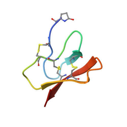 | |
UniProt | |||||
Entity Groups | |||||
| Sequence Clusters | 30% Identity50% Identity70% Identity90% Identity95% Identity100% Identity | ||||
| UniProt Group | I1SB10 | ||||
Sequence AnnotationsExpand | |||||
Reference Sequence | |||||
| Ligands 1 Unique | |||||
|---|---|---|---|---|---|
| ID | Chains | Name / Formula / InChI Key | 2D Diagram | 3D Interactions | |
| MOH Download:Ideal Coordinates CCD File | B [auth A] C [auth A] D [auth A] E [auth A] F [auth A] | METHANOL C H4 O OKKJLVBELUTLKV-UHFFFAOYSA-N |  | ||
| Modified Residues 1 Unique | |||||
|---|---|---|---|---|---|
| ID | Chains | Type | Formula | 2D Diagram | Parent |
| PCA Query on PCA | A | L-PEPTIDE LINKING | C5 H7 N O3 |  | GLN |
| Length ( Å ) | Angle ( ˚ ) |
|---|---|
| a = 14.8028 | α = 83.19 |
| b = 18.5573 | β = 84.1 |
| c = 24.1784 | γ = 68.04 |
| Software Name | Purpose |
|---|---|
| SHELX | refinement |
| PDB_EXTRACT | data extraction |
| SHELXL | refinement |
| SHELXL-97 | refinement |


