3Q2C | pdb_00003q2c

Binding properties to HLA class I molecules and the structure of the leukocyte Ig-like receptor A3 (LILRA3/ILT6/LIR4/CD85e)


Experimental Data Snapshot

  • Method: X-RAY DIFFRACTION
  • Resolution: 2.50 Å
  • R-Value Free: 
    0.257 (Depositor), 0.264 (DCC) 
  • R-Value Work: 
    0.225 (Depositor), 0.229 (DCC) 
  • R-Value Observed: 
    0.226 (Depositor) 

Starting Model: experimental
View more details

wwPDB Validation 3D Report Full Report

Validation slider image for 3Q2C

This is version 1.2 of the entry. See complete history

Literature

LILRA3 binds both classical and non-classical HLA class I molecules but with reduced affinities compared to LILRB1/LILRB2: structural evidence

Ryu, M.Chen, Y.Qi, J.Liu, J.Fan, Z.Nam, G.Shi, Y.Cheng, H.Gao, G.F.

(2011) PLoS One 6: e19245-e19245

  • DOI: https://doi.org/10.1371/journal.pone.0019245
  • Primary Citation Related Structures: 
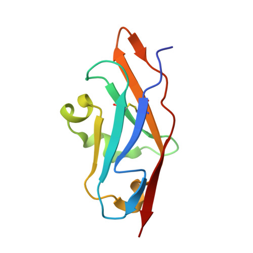
    3Q2C

  • PubMed Abstract: 

    Structurally, Group 1 LILR (Leukocyte Immunoglobulin (Ig)-Like Receptor, also known as Ig-like transcripts, ILT; Leukocyte Ig-like receptor, LIR; and CD85) members are very similar in terms of the HLAIs (human leukocyte antigen class I molecules) binding region and were hypothesized that they all bind to HLAIs. As one of the Group 1 LILRs, LILRA3 is the only secretory LILR and may greatly control the inhibitory immune response induced by LILRB1, LILRB2, and other HLA-binding LILR molecules like LILRA1. Nevertheless, little was known about the binding of LILRA3 to HLAIs. In this report, we present the crystal structure of the LILRA3 domain 1 (D1) and evaluate the D1 and D1D2 (domain 1 and domain 2) binding to classical and non-classical HLAIs using BIAcore® surface plasmon resonance analysis (SPR). We found that LILRA3 binds both classical HLA-A*0201 and non-classical HLA-G1 but with reduced affinities compared to either LILRB1 or LILRB2. The polymorphic amino acids and the LILRA3 D1 structure support this notion.


  • Organizational Affiliation
    • CAS Key Laboratory of Pathogenic Microbiology and Immunology, Institute of Microbiology, Chinese Academy of Sciences, Beijing, China.

Macromolecule Content 

  • Total Structure Weight: 10.79 kDa 
  • Atom Count: 787 
  • Modeled Residue Count: 96 
  • Deposited Residue Count: 97 
  • Unique protein chains: 1

Macromolecules

Find similar proteins by:|  3D Structure
Entity ID: 1
MoleculeChains  Sequence LengthOrganismDetailsImage
Leukocyte immunoglobulin-like receptor subfamily A member 397Homo sapiensMutation(s): 0 
UniProt
Find proteins for Q8N6C8 (Homo sapiens)
Explore Q8N6C8 
Go to UniProtKB:  Q8N6C8
Entity Groups
Sequence Clusters30% Identity50% Identity70% Identity90% Identity95% Identity100% Identity
UniProt GroupQ8N6C8
Sequence Annotations
Expand
Reference Sequence

Experimental Data & Validation

Experimental Data

  • Method: X-RAY DIFFRACTION
  • Resolution: 2.50 Å
  • R-Value Free:  0.257 (Depositor), 0.264 (DCC) 
  • R-Value Work:  0.225 (Depositor), 0.229 (DCC) 
  • R-Value Observed: 0.226 (Depositor) 
Space Group: C 1 2 1
Unit Cell:
Length ( Å )Angle ( ˚ )
a = 83.308α = 90
b = 27.817β = 111.65
c = 48.257γ = 90
Software Package:
Software NamePurpose
CrystalCleardata collection
PHASERphasing
PHENIXrefinement
HKL-2000data reduction
HKL-2000data scaling

Structure Validation

View Full Validation Report


Entry History 

Deposition Data

Revision History  (Full details and data files)

  • Version 1.0: 2011-07-13
    Type: Initial release
  • Version 1.1: 2023-11-01
    Changes: Data collection, Database references, Refinement description
  • Version 1.2: 2024-10-30
    Changes: Structure summary