3Q25 | pdb_00003q25

Crystal structure of human alpha-synuclein (1-19) fused to maltose binding protein (MBP)


Experimental Data Snapshot

  • Method: X-RAY DIFFRACTION
  • Resolution: 1.90 Å
  • R-Value Free: 
    0.196 (Depositor), 0.197 (DCC) 
  • R-Value Work: 
    0.170 (Depositor), 0.169 (DCC) 
  • R-Value Observed: 
    0.171 (Depositor) 

Starting Model: experimental
View more details

wwPDB Validation 3D Report Full Report

Validation slider image for 3Q25

This is version 2.1 of the entry. See complete history

Literature

Structures of segments of alpha-synuclein fused to maltose-binding protein suggest intermediate states during amyloid formation

Zhao, M.Cascio, D.Sawaya, M.R.Eisenberg, D.

(2011) Protein Sci 20: 996-1004

  • DOI: https://doi.org/10.1002/pro.630
  • Primary Citation Related Structures: 
    3Q25, 3Q26, 3Q27, 3Q28, 3Q29

  • PubMed Abstract: 

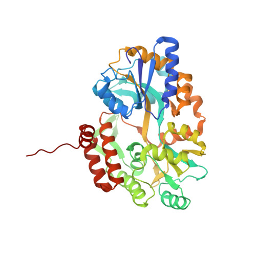
    Aggregates of the protein α-synuclein are the main component of Lewy bodies, the hallmark of Parkinson's disease. α-Synuclein aggregates are also found in many human neurodegenerative diseases known as synucleinopathies. In vivo, α-synuclein associates with membranes and adopts α-helical conformations. The details of how α-synuclein converts from the functional native state to amyloid aggregates remain unknown. In this study, we use maltose-binding protein (MBP) as a carrier to crystallize segments of α-synuclein. From crystal structures of fusions between MBP and four segments of α-synuclein, we have been able to trace a virtual model of the first 72 residues of α-synuclein. Instead of a mostly α-helical conformation observed in the lipid environment, our crystal structures show α-helices only at residues 1-13 and 20-34. The remaining segments are extended loops or coils. All of the predicted fiber-forming segments based on the 3D profile method are in extended conformations. We further show that the MBP fusion proteins with fiber-forming segments from α-synuclein can also form fiber-like nano-crystals or amyloid-like fibrils. Our structures suggest intermediate states during amyloid formation of α-synuclein.


  • Organizational Affiliation
    • Department of Biological Chemistry, David Geffen School of Medicine, University of California, Los Angeles, California 90095, USA.

Macromolecule Content 

  • Total Structure Weight: 44.55 kDa 
  • Atom Count: 3,392 
  • Modeled Residue Count: 388 
  • Deposited Residue Count: 390 
  • Unique protein chains: 1

Macromolecules

Find similar proteins by:|  3D Structure
Entity ID: 1
MoleculeChains  Sequence LengthOrganismDetailsImage
Maltose-binding periplasmic protein/alpha-synuclein chimeric protein390Escherichia coliHomo sapiens
This entity is chimeric
Mutation(s): 0 
UniProt & NIH Common Fund Data Resources
Find proteins for P0AEX9 (Escherichia coli (strain K12))
Explore P0AEX9 
Go to UniProtKB:  P0AEX9
Find proteins for P37840 (Homo sapiens)
Explore P37840 
Go to UniProtKB:  P37840
PHAROS:  P37840
GTEx:  ENSG00000145335 
Entity Groups
Sequence Clusters30% Identity50% Identity70% Identity90% Identity95% Identity100% Identity
UniProt GroupsP37840P0AEX9
Sequence Annotations
Expand
Reference Sequence

Oligosaccharides

Help  
Entity ID: 2
MoleculeChains Length2D Diagram GlycosylationD Interactions
alpha-D-glucopyranose-(1-4)-alpha-D-glucopyranose
B
2N/A
Glycosylation Resources
GlyTouCan: G07411ON
GlyCosmos: G07411ON

Small Molecules

Ligands 2 Unique
IDChains Name / Formula / InChI Key2D Diagram3D Interactions
SO4

Query on SO4


Download:Ideal Coordinates CCD File
C [auth A]
D [auth A]
E [auth A]
F [auth A]
G [auth A]
C [auth A],
D [auth A],
E [auth A],
F [auth A],
G [auth A],
H [auth A],
I [auth A],
J [auth A],
K [auth A],
L [auth A]
SULFATE ION
O4 S
QAOWNCQODCNURD-UHFFFAOYSA-L
GOL

Query on GOL


Download:Ideal Coordinates CCD File
M [auth A]
N [auth A]
O [auth A]
P [auth A]
Q [auth A]
M [auth A],
N [auth A],
O [auth A],
P [auth A],
Q [auth A],
R [auth A]
GLYCEROL
C3 H8 O3
PEDCQBHIVMGVHV-UHFFFAOYSA-N

Biologically Interesting Molecules (External Reference) 

1 Unique

Experimental Data & Validation

Experimental Data

  • Method: X-RAY DIFFRACTION
  • Resolution: 1.90 Å
  • R-Value Free:  0.196 (Depositor), 0.197 (DCC) 
  • R-Value Work:  0.170 (Depositor), 0.169 (DCC) 
  • R-Value Observed: 0.171 (Depositor) 
Space Group: P 43 21 2
Unit Cell:
Length ( Å )Angle ( ˚ )
a = 77.47α = 90
b = 77.47β = 90
c = 172.64γ = 90
Software Package:
Software NamePurpose
XSCALEdata scaling
PHASERphasing
PHENIXrefinement
PDB_EXTRACTdata extraction
XDSdata reduction

Structure Validation

View Full Validation Report


Entry History 

Deposition Data

Revision History  (Full details and data files)

  • Version 1.0: 2011-06-01
    Type: Initial release
  • Version 1.1: 2011-07-13
    Changes: Version format compliance
  • Version 1.2: 2017-08-02
    Changes: Refinement description, Source and taxonomy
  • Version 2.0: 2020-07-29
    Type: Remediation
    Reason: Carbohydrate remediation
    Changes: Atomic model, Data collection, Database references, Derived calculations, Non-polymer description, Structure summary
  • Version 2.1: 2023-09-13
    Changes: Data collection, Database references, Refinement description, Structure summary