3Q13 | pdb_00003q13

The Structure of the Ca2+-binding, Glycosylated F-spondin Domain of F-spondin, A C2-domain Variant from Extracellular Matrix


Experimental Data Snapshot

  • Method: X-RAY DIFFRACTION
  • Resolution: 1.95 Å
  • R-Value Free: 
    0.210 (Depositor), 0.213 (DCC) 
  • R-Value Work: 
    0.167 (Depositor), 0.167 (DCC) 
  • R-Value Observed: 
    0.169 (Depositor) 

Starting Model: experimental
View more details

wwPDB Validation 3D Report Full Report

Validation slider image for 3Q13

This is version 2.2 of the entry. See complete history

Literature

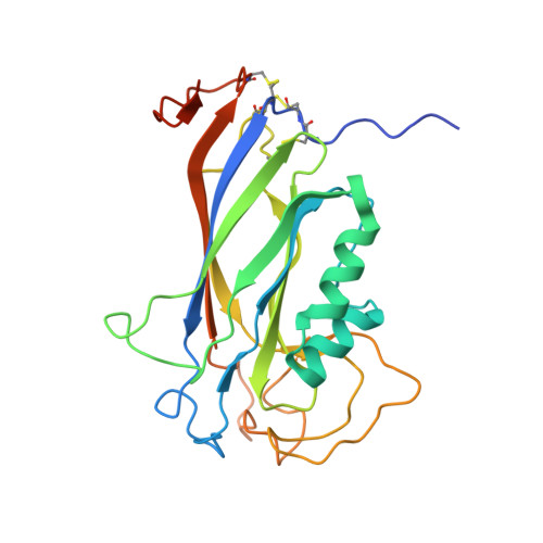
The structure of the Ca2+-binding, glycosylated F-spondin domain of F-spondin - A C2-domain variant in an extracellular matrix protein.

Tan, K.Lawler, J.

(2011) BMC Struct Biol 11: 22-22

  • DOI: https://doi.org/10.1186/1472-6807-11-22
  • Primary Citation Related Structures: 
    3Q13

  • PubMed Abstract: 

    F-spondin is a multi-domain extracellular matrix (ECM) protein and a contact-repellent molecule that directs axon outgrowth and cell migration during development. The reelin_N domain and the F-spondin domain (FS domain) comprise a proteolytic fragment that interacts with the cell membrane and guides the projection of commissural axons to floor plate. The FS domain is found in F-spondins, mindins, M-spondin and amphiF-spondin. We present the crystal structure of human F-spondin FS domain at 1.95Å resolution. The structure reveals a Ca2+-binding C2 domain variant with an 8-stranded antiparallel β-sandwich fold. Though the primary sequences of the FS domains of F-spondin and mindin are less than 36% identical, their overall structures are very similar. The unique feature of F-spondin FS domain is the presence of three disulfide bonds associated with the N- and C-termini of the domain and a highly conserved N-linked glycosylation site. The integrin-binding motif found in mindin is not conserved in the F-spondin FS domain. The structure of the F-spondin FS domain completes the structural studies of the multiple-domain ECM molecule. The homology of its core structure to a common Ca2+- and lipid-binding C2 domain suggests that the F-spondin FS domain may be responsible for part of the membrane targeting of F-spondin in its regulation of axon development. The structural properties of the FS domain revealed in this study pave the way for further exploration into the functions of F-spondin.


  • Organizational Affiliation
    • Midwest Center for Structural Genomics and Structural Biology Center, Biosciences Division, Argonne National Laboratory, Argonne, IL 60439, USA. ktan@anl.gov

Macromolecule Content 

  • Total Structure Weight: 29.9 kDa 
  • Atom Count: 2,314 
  • Modeled Residue Count: 236 
  • Deposited Residue Count: 258 
  • Unique protein chains: 1

Macromolecules

Find similar proteins by:|  3D Structure
Entity ID: 1
MoleculeChains  Sequence LengthOrganismDetailsImage
Spondin-1258Homo sapiensMutation(s): 0 
Gene Names: Homo sapiensKIAA0762SPON1VSGP
UniProt & NIH Common Fund Data Resources
Find proteins for Q9HCB6 (Homo sapiens)
Explore Q9HCB6 
Go to UniProtKB:  Q9HCB6
PHAROS:  Q9HCB6
GTEx:  ENSG00000262655 
Entity Groups
Sequence Clusters30% Identity50% Identity70% Identity90% Identity95% Identity100% Identity
UniProt GroupQ9HCB6
Glycosylation
Glycosylation Sites: 1Go to GlyGen: Q9HCB6-1
Sequence Annotations
Expand
Reference Sequence

Oligosaccharides

Help  
Entity ID: 2
MoleculeChains Length2D Diagram GlycosylationD Interactions
beta-D-mannopyranose-(1-4)-2-acetamido-2-deoxy-beta-D-glucopyranose-(1-4)-2-acetamido-2-deoxy-beta-D-glucopyranose
B
3N-Glycosylation
Glycosylation Resources
GlyTouCan: G15407YE
GlyCosmos: G15407YE
GlyGen: G15407YE

Experimental Data & Validation

Experimental Data

  • Method: X-RAY DIFFRACTION
  • Resolution: 1.95 Å
  • R-Value Free:  0.210 (Depositor), 0.213 (DCC) 
  • R-Value Work:  0.167 (Depositor), 0.167 (DCC) 
  • R-Value Observed: 0.169 (Depositor) 
Space Group: P 6 2 2
Unit Cell:
Length ( Å )Angle ( ˚ )
a = 120.244α = 90
b = 120.244β = 90
c = 86.362γ = 120
Software Package:
Software NamePurpose
SBC-Collectdata collection
MOLREPphasing
REFMACrefinement
HKL-3000data reduction
HKL-3000data scaling

Structure Validation

View Full Validation Report


Entry History 

Deposition Data

Revision History  (Full details and data files)

  • Version 1.0: 2011-06-29
    Type: Initial release
  • Version 1.1: 2011-07-13
    Changes: Version format compliance
  • Version 2.0: 2020-07-29
    Type: Remediation
    Reason: Carbohydrate remediation
    Changes: Atomic model, Data collection, Database references, Derived calculations, Structure summary
  • Version 2.1: 2023-09-13
    Changes: Data collection, Database references, Refinement description, Structure summary
  • Version 2.2: 2024-11-27
    Changes: Structure summary