A metal switch for controlling the activity of molecular motor proteins.
Cochran, J.C., Zhao, Y.C., Wilcox, D.E., Kull, F.J.(2012) Nat Struct Mol Biol 19: 122-127
- PubMed: 22198464 Search on PubMedSearch on PubMed Central
- DOI: https://doi.org/10.1038/nsmb.2190
- Primary Citation Related Structures:
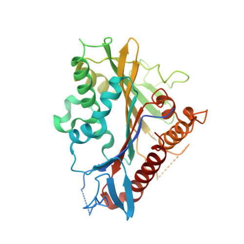
3PXN - PubMed Abstract:
Kinesins are molecular motors that require a divalent metal ion (for example, Mg(2+)) to convert the energy of ATP hydrolysis into directed force production along microtubules. Here we present the crystal structure of a recombinant kinesin motor domain bound to Mn(2+) and ADP and report on a serine-to-cysteine substitution in the switch 1 motif of kinesin that allows its ATP hydrolysis activity to be controlled by adjusting the ratio of Mn(2+) to Mg(2+). This mutant kinesin binds ATP similarly in the presence of either metal ion, but its ATP hydrolysis activity is greatly diminished in the presence of Mg(2+). In human kinesin-1 and kinesin-5 as well as Drosophila melanogaster kinesin-10 and kinesin-14, this defect is rescued by Mn(2+), providing a way to control both the enzymatic activity and force-generating ability of these nanomachines.
- Dartmouth College, Department of Chemistry, Hanover, New Hampshire, USA.
Organizational Affiliation: