3PSY | pdb_00003psy

Endothiapepsin in complex with an inhibitor based on the Gewald reaction


Experimental Data Snapshot

  • Method: X-RAY DIFFRACTION
  • Resolution: 1.43 Å
  • R-Value Free: 
    0.167 (Depositor), 0.172 (DCC) 
  • R-Value Work: 
    0.128 (Depositor), 0.136 (DCC) 

Starting Model: experimental
View more details

wwPDB Validation 3D Report Full Report

Validation slider image for 3PSY

Ligand Structure Quality Assessment 


This is version 1.3 of the entry. See complete history

Literature

Tracing binding modes in hit-to-lead optimization: chameleon-like poses of aspartic protease inhibitors.

Kuhnert, M.Koster, H.Bartholomaus, R.Park, A.Y.Shahim, A.Heine, A.Steuber, H.Klebe, G.Diederich, W.E.

(2015) Angew Chem Int Ed Engl 54: 2849-2853

  • DOI: https://doi.org/10.1002/anie.201411206
  • Primary Citation Related Structures: 
    3PSY, 3WZ6, 3WZ7, 3WZ8

  • PubMed Abstract: 

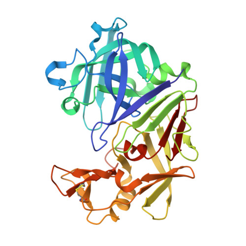
    Successful lead optimization in structure-based drug discovery depends on the correct deduction and interpretation of the underlying structure-activity relationships (SAR) to facilitate efficient decision-making on the next candidates to be synthesized. Consequently, the question arises, how frequently a binding mode (re)-validation is required, to ensure not to be misled by invalid assumptions on the binding geometry. We present an example in which minor chemical modifications within one inhibitor series lead to surprisingly different binding modes. X-ray structure determination of eight inhibitors derived from one core scaffold resulted in four different binding modes in the aspartic protease endothiapepsin, a well-established surrogate for e.g. renin and β-secretase. In addition, we suggest an empirical metrics that might serve as an indicator during lead optimization to qualify compounds as candidates for structural revalidation.


  • Organizational Affiliation
    • Institut für Pharmazeutische Chemie, Philipps-Universität Marburg, Hans-Meerwein-Strasse 3, 35032 Marburg (Germany).

Macromolecule Content 

  • Total Structure Weight: 34.93 kDa 
  • Atom Count: 2,705 
  • Modeled Residue Count: 330 
  • Deposited Residue Count: 330 
  • Unique protein chains: 1

Macromolecules

Find similar proteins by:|  3D Structure
Entity ID: 1
MoleculeChains  Sequence LengthOrganismDetailsImage
Endothiapepsin330Cryphonectria parasiticaMutation(s): 0 
EC: 3.4.23.22
UniProt
Find proteins for P11838 (Cryphonectria parasitica)
Explore P11838 
Go to UniProtKB:  P11838
Entity Groups
Sequence Clusters30% Identity50% Identity70% Identity90% Identity95% Identity100% Identity
UniProt GroupP11838
Sequence Annotations
Expand
Reference Sequence

Small Molecules

Ligands 5 Unique
IDChains Name / Formula / InChI Key2D Diagram3D Interactions
RB9

Query on RB9


Download:Ideal Coordinates CCD File
B [auth A]N-benzyl-2-({N-[2-(1H-indol-3-yl)ethyl]glycyl}amino)-4-phenylthiophene-3-carboxamide
C30 H28 N4 O2 S
NZMGUKNNKODUOD-UHFFFAOYSA-N
1PE

Query on 1PE


Download:Ideal Coordinates CCD File
F [auth A]PENTAETHYLENE GLYCOL
C10 H22 O6
JLFNLZLINWHATN-UHFFFAOYSA-N
PG4

Query on PG4


Download:Ideal Coordinates CCD File
E [auth A]TETRAETHYLENE GLYCOL
C8 H18 O5
UWHCKJMYHZGTIT-UHFFFAOYSA-N
GOL

Query on GOL


Download:Ideal Coordinates CCD File
D [auth A]GLYCEROL
C3 H8 O3
PEDCQBHIVMGVHV-UHFFFAOYSA-N
DMS

Query on DMS


Download:Ideal Coordinates CCD File
C [auth A]DIMETHYL SULFOXIDE
C2 H6 O S
IAZDPXIOMUYVGZ-UHFFFAOYSA-N

Experimental Data & Validation

Experimental Data

  • Method: X-RAY DIFFRACTION
  • Resolution: 1.43 Å
  • R-Value Free:  0.167 (Depositor), 0.172 (DCC) 
  • R-Value Work:  0.128 (Depositor), 0.136 (DCC) 
Space Group: P 1 21 1
Unit Cell:
Length ( Å )Angle ( ˚ )
a = 45.45α = 90
b = 73.05β = 109.5
c = 52.78γ = 90
Software Package:
Software NamePurpose
MAR345dtbdata collection
PHASERphasing
SHELXL-97refinement
HKL-2000data reduction
HKL-2000data scaling

Structure Validation

View Full Validation Report


Ligand Structure Quality Assessment 


Entry History 

Deposition Data

Revision History  (Full details and data files)

  • Version 1.0: 2011-12-07
    Type: Initial release
  • Version 1.1: 2019-12-11
    Changes: Data collection, Database references
  • Version 1.2: 2023-11-01
    Changes: Data collection, Database references, Derived calculations, Refinement description
  • Version 1.3: 2024-11-13
    Changes: Structure summary