3PPM | pdb_00003ppm

Crystal Structure of a Noncovalently Bound alpha-Ketoheterocycle Inhibitor (Phenhexyl/Oxadiazole/Pyridine) to a Humanized Variant of Fatty Acid Amide Hydrolase


Experimental Data Snapshot

  • Method: X-RAY DIFFRACTION
  • Resolution: 1.78 Å
  • R-Value Free: 
    0.176 (Depositor), 0.173 (DCC) 
  • R-Value Work: 
    0.151 (Depositor), 0.149 (DCC) 
  • R-Value Observed: 
    0.152 (Depositor) 

Starting Model: experimental
View more details

wwPDB Validation 3D Report Full Report

Validation slider image for 3PPM

Ligand Structure Quality Assessment 


This is version 1.1 of the entry. See complete history

Literature

Fluoride-mediated capture of a noncovalent bound state of a reversible covalent enzyme inhibitor: X-ray crystallographic analysis of an exceptionally potent alpha-ketoheterocycle inhibitor of fatty acid amide hydrolase.

Mileni, M.Garfunkle, J.Ezzili, C.Cravatt, B.F.Stevens, R.C.Boger, D.L.

(2011) J Am Chem Soc 133: 4092-4100

  • DOI: https://doi.org/10.1021/ja110877y
  • Primary Citation Related Structures: 
    3PPM, 3PR0

  • PubMed Abstract: 

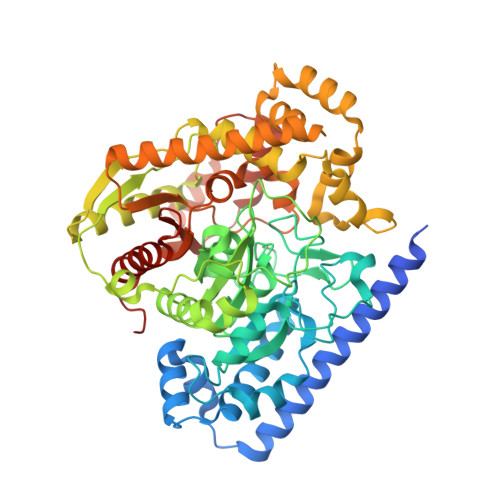
    Two cocrystal X-ray structures of the exceptionally potent α-ketoheterocycle inhibitor 1 (K(i) = 290 pM) bound to a humanized variant of rat fatty acid amide hydrolase (FAAH) are disclosed, representing noncovalently and covalently bound states of the same inhibitor with the enzyme. Key to securing the structure of the noncovalently bound state of the inhibitor was the inclusion of fluoride ion in the crystallization conditions that is proposed to bind the oxyanion hole precluding inhibitor covalent adduct formation with stabilization of the tetrahedral hemiketal. This permitted the opportunity to detect important noncovalent interactions stabilizing the binding of the inhibitor within the FAAH active site independent of the covalent reaction. Remarkably, noncovalently bound 1 in the presence of fluoride appears to capture the active site in the same "in action" state with the three catalytic residues Ser241-Ser217-Lys142 occupying essentially identical positions observed in the covalently bound structure of 1, suggesting that this technique of introducing fluoride may have important applications in structural studies beyond inhibiting substrate or inhibitor oxyanion hole binding. Key insights to emerge from the studies include the observations that noncovalently bound 1 binds in its ketone (not gem diol) form, that the terminal phenyl group in the acyl side chain of the inhibitor serves as the key anchoring interaction overriding the intricate polar interactions in the cytosolic port, and that the role of the central activating heterocycle is dominated by its intrinsic electron-withdrawing properties. These two structures are also briefly compared with five X-ray structures of α-ketoheterocycle-based inhibitors bound to FAAH recently disclosed.


  • Organizational Affiliation
    • Department of Chemistry, Scripps Research Institute, 10550 North Torrey Pines Road, La Jolla, California 92037, United States.

Macromolecule Content 

  • Total Structure Weight: 127.65 kDa 
  • Atom Count: 9,708 
  • Modeled Residue Count: 1,091 
  • Deposited Residue Count: 1,146 
  • Unique protein chains: 1

Macromolecules

Find similar proteins by:|  3D Structure
Entity ID: 1
MoleculeChains  Sequence LengthOrganismDetailsImage
Fatty-acid amide hydrolase 1
A, B
573Rattus norvegicusMutation(s): 6 
Gene Names: Faahfaah-1Faah1
EC: 3.5.1.99 (PDB Primary Data), 3.1.1 (UniProt)
UniProt
Find proteins for P97612 (Rattus norvegicus)
Explore P97612 
Go to UniProtKB:  P97612
Entity Groups
Sequence Clusters30% Identity50% Identity70% Identity90% Identity95% Identity100% Identity
UniProt GroupP97612
Sequence Annotations
Expand
Reference Sequence

Small Molecules

Ligands 5 Unique
IDChains Name / Formula / InChI Key2D Diagram3D Interactions
JG1

Query on JG1


Download:Ideal Coordinates CCD File
C [auth A],
I [auth B]
7-phenyl-1-[5-(pyridin-2-yl)-1,3,4-oxadiazol-2-yl]heptan-1-one
C20 H21 N3 O2
BYQFWIOGPFHXFV-UHFFFAOYSA-N
1DO

Query on 1DO


Download:Ideal Coordinates CCD File
L [auth B]1-DODECANOL
C12 H26 O
LQZZUXJYWNFBMV-UHFFFAOYSA-N
PEG

Query on PEG


Download:Ideal Coordinates CCD File
G [auth A],
H [auth A],
M [auth B],
N [auth B]
DI(HYDROXYETHYL)ETHER
C4 H10 O3
MTHSVFCYNBDYFN-UHFFFAOYSA-N
CL

Query on CL


Download:Ideal Coordinates CCD File
D [auth A],
E [auth A],
J [auth B]
CHLORIDE ION
Cl
VEXZGXHMUGYJMC-UHFFFAOYSA-M
F

Query on F


Download:Ideal Coordinates CCD File
F [auth A],
K [auth B]
FLUORIDE ION
F
KRHYYFGTRYWZRS-UHFFFAOYSA-M

Experimental Data & Validation

Experimental Data

  • Method: X-RAY DIFFRACTION
  • Resolution: 1.78 Å
  • R-Value Free:  0.176 (Depositor), 0.173 (DCC) 
  • R-Value Work:  0.151 (Depositor), 0.149 (DCC) 
  • R-Value Observed: 0.152 (Depositor) 
Space Group: P 32 2 1
Unit Cell:
Length ( Å )Angle ( ˚ )
a = 103.692α = 90
b = 103.692β = 90
c = 254.705γ = 120
Software Package:
Software NamePurpose
HKL-2000data collection
PHASERphasing
PHENIXrefinement
HKL-2000data reduction
HKL-2000data scaling

Structure Validation

View Full Validation Report


Ligand Structure Quality Assessment 


Entry History 

Deposition Data

Revision History  (Full details and data files)

  • Version 1.0: 2011-11-09
    Type: Initial release
  • Version 1.1: 2023-09-06
    Changes: Data collection, Database references, Derived calculations, Refinement description