3PHM | pdb_00003phm

REDUCED (CU+) PEPTIDYLGLYCINE ALPHA-HYDROXYLATING MONOOXYGENASE (PHM)


Experimental Data Snapshot

  • Method: X-RAY DIFFRACTION
  • Resolution: 2.10 Å
  • R-Value Free: 
    0.257 (Depositor) 
  • R-Value Work: 
    0.200 (Depositor) 

Starting Model: experimental
View more details

wwPDB Validation 3D Report Full Report

Validation slider image for 3PHM

This is version 1.4 of the entry. See complete history

Literature

Substrate-mediated electron transfer in peptidylglycine alpha-hydroxylating monooxygenase.

Prigge, S.T.Kolhekar, A.S.Eipper, B.A.Mains, R.E.Amzel, L.M.

(1999) Nat Struct Biol 6: 976-983

  • DOI: https://doi.org/10.1038/13351
  • Primary Citation Related Structures: 
    1OPM, 3PHM

  • PubMed Abstract: 

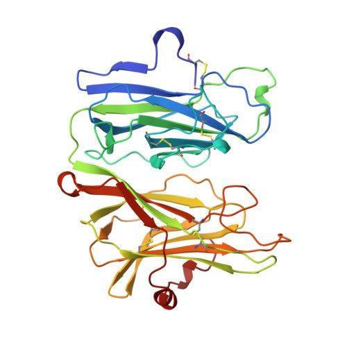
    Peptide amidation is a ubiquitous posttranslational modification of bioactive peptides. Peptidylglycine alpha-hydroxylating monooxygenase (PHM; EC 1.14.17.3), the enzyme that catalyzes the first step of this reaction, is composed of two domains, each of which binds one copper atom. The coppers are held 11 A apart on either side of a solvent-filled interdomain cleft, and the PHM reaction requires electron transfer between these sites. A plausible mechanism for electron transfer might involve interdomain motion to decrease the distance between the copper atoms. Our experiments show that PHM catalytic core (PHMcc) is enzymatically active in the crystal phase, where interdomain motion is not possible. Instead, structures of two states relevant to catalysis indicate that water, substrate and active site residues may provide an electron transfer pathway that exists only during the PHM catalytic cycle.


  • Organizational Affiliation
    • Department of Biophysics and Biophysical Chemistry, Johns Hopkins School of Medicine, Baltimore, Maryland 21205, USA.

Macromolecule Content 

  • Total Structure Weight: 35.27 kDa 
  • Atom Count: 2,622 
  • Modeled Residue Count: 310 
  • Deposited Residue Count: 310 
  • Unique protein chains: 1

Macromolecules

Find similar proteins by:|  3D Structure
Entity ID: 1
MoleculeChains  Sequence LengthOrganismDetailsImage
PROTEIN (PEPTIDYLGLYCINE ALPHA-HYDROXYLATING MONOOXYGENASE)310Rattus norvegicusMutation(s): 0 
EC: 1.14.17.3 (PDB Primary Data), 4.3.2.5 (UniProt)
UniProt
Find proteins for P14925 (Rattus norvegicus)
Explore P14925 
Go to UniProtKB:  P14925
Entity Groups
Sequence Clusters30% Identity50% Identity70% Identity90% Identity95% Identity100% Identity
UniProt GroupP14925
Sequence Annotations
Expand
Reference Sequence

Small Molecules

Ligands 4 Unique
IDChains Name / Formula / InChI Key2D Diagram3D Interactions
GOL

Query on GOL


Download:Ideal Coordinates CCD File
F [auth A],
G [auth A],
H [auth A],
I [auth A],
J [auth A]
GLYCEROL
C3 H8 O3
PEDCQBHIVMGVHV-UHFFFAOYSA-N
CU

Query on CU


Download:Ideal Coordinates CCD File
B [auth A],
D [auth A]
COPPER (II) ION
Cu
JPVYNHNXODAKFH-UHFFFAOYSA-N
NI

Query on NI


Download:Ideal Coordinates CCD File
E [auth A]NICKEL (II) ION
Ni
VEQPNABPJHWNSG-UHFFFAOYSA-N
AZI

Query on AZI


Download:Ideal Coordinates CCD File
C [auth A]AZIDE ION
N3
IVRMZWNICZWHMI-UHFFFAOYSA-N

Experimental Data & Validation

Experimental Data

  • Method: X-RAY DIFFRACTION
  • Resolution: 2.10 Å
  • R-Value Free:  0.257 (Depositor) 
  • R-Value Work:  0.200 (Depositor) 
Space Group: P 21 21 21
Unit Cell:
Length ( Å )Angle ( ˚ )
a = 69.519α = 90
b = 68.77β = 90
c = 82.417γ = 90
Software Package:
Software NamePurpose
DENZOdata reduction
SCALEPACKdata scaling
X-PLORrefinement

Structure Validation

View Full Validation Report


Entry History 

Deposition Data

Revision History  (Full details and data files)

  • Version 1.0: 1999-09-29
    Type: Initial release
  • Version 1.1: 2007-10-16
    Changes: Version format compliance
  • Version 1.2: 2011-07-13
    Changes: Non-polymer description, Version format compliance
  • Version 1.3: 2023-09-06
    Changes: Data collection, Database references, Derived calculations, Refinement description
  • Version 1.4: 2024-10-30
    Changes: Structure summary