3PGK | pdb_00003pgk

The structure of yeast phosphoglycerate kinase at 0.25 nm resolution


Experimental Data Snapshot

  • Method: X-RAY DIFFRACTION
  • Resolution: 2.50 Å

wwPDB Validation 3D Report Full Report

Validation slider image for 3PGK

This is version 1.5 of the entry. See complete history

Literature

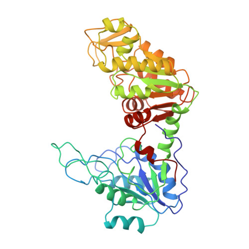
Sequence and structure of yeast phosphoglycerate kinase.

Watson, H.C.Walker, N.P.Shaw, P.J.Bryant, T.N.Wendell, P.L.Fothergill, L.A.Perkins, R.E.Conroy, S.C.Dobson, M.J.Tuite, M.F.

(1982) EMBO J 1: 1635-1640

  • DOI: https://doi.org/10.1002/j.1460-2075.1982.tb01366.x
  • Primary Citation Related Structures: 
    3PGK

  • PubMed Abstract: 

    The structure of yeast phosphoglycerate kinase has been determined with data obtained from amino acid sequence, nucleotide sequence, and X-ray crystallographic studies. The substrate binding sites, as deduced from electron density maps, are compatible with known substrate specificity and the stereochemical requirements for the enzymic reaction. A carboxyl-imidazole interaction appears to be involved in controlling the transition between the open and closed forms of the enzyme.

Macromolecule Content 

  • Total Structure Weight: 45.37 kDa 
  • Atom Count: 3,191 
  • Modeled Residue Count: 416 
  • Deposited Residue Count: 416 
  • Unique protein chains: 1

Macromolecules

Find similar proteins by:|  3D Structure
Entity ID: 1
MoleculeChains  Sequence LengthOrganismDetailsImage
PHOSPHOGLYCERATE KINASE416Saccharomyces cerevisiaeMutation(s): 0 
EC: 2.7.2.3
UniProt
Find proteins for P00560 (Saccharomyces cerevisiae (strain ATCC 204508 / S288c))
Explore P00560 
Go to UniProtKB:  P00560
Entity Groups
Sequence Clusters30% Identity50% Identity70% Identity90% Identity95% Identity100% Identity
UniProt GroupP00560
Sequence Annotations
Expand
Reference Sequence

Small Molecules

Ligands 3 Unique
IDChains Name / Formula / InChI Key2D Diagram3D Interactions
ATP

Query on ATP


Download:Ideal Coordinates CCD File
C [auth A]ADENOSINE-5'-TRIPHOSPHATE
C10 H16 N5 O13 P3
ZKHQWZAMYRWXGA-KQYNXXCUSA-N
3PG

Query on 3PG


Download:Ideal Coordinates CCD File
D [auth A]3-PHOSPHOGLYCERIC ACID
C3 H7 O7 P
OSJPPGNTCRNQQC-UWTATZPHSA-N
MG

Query on MG


Download:Ideal Coordinates CCD File
B [auth A]MAGNESIUM ION
Mg
JLVVSXFLKOJNIY-UHFFFAOYSA-N

Experimental Data & Validation

Experimental Data

  • Method: X-RAY DIFFRACTION
  • Resolution: 2.50 Å
Space Group: C 1 2 1
Unit Cell:
Length ( Å )Angle ( ˚ )
a = 126.6α = 90
b = 54.4β = 134.4
c = 93γ = 90

Structure Validation

View Full Validation Report


Entry History 

Deposition Data

Revision History  (Full details and data files)

  • Version 1.0: 1982-09-24
    Type: Initial release
  • Version 1.1: 2008-03-25
    Changes: Version format compliance
  • Version 1.2: 2011-07-13
    Changes: Version format compliance
  • Version 1.3: 2017-11-29
    Changes: Derived calculations, Other
  • Version 1.4: 2024-02-21
    Changes: Data collection, Database references, Derived calculations
  • Version 1.5: 2024-11-20
    Changes: Structure summary