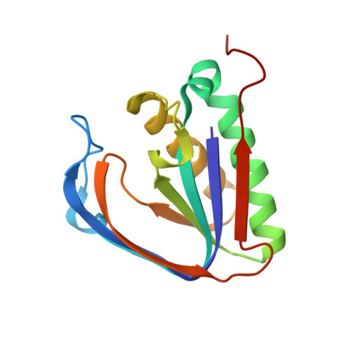
3PD4

Crystal structure of the editing domain of threonyl-tRNA synthetase from Pyrococcus abyssi in complex with glycyl-3'-aminoadenosine


Experimental Data Snapshot

  • Method: X-RAY DIFFRACTION
  • Resolution: 2.40 Å
  • R-Value Free: 0.289 
  • R-Value Work: 0.204 
  • R-Value Observed: 0.208 

Starting Model: experimental
View more details

wwPDB Validation   3D Report Full Report


Ligand Structure Quality Assessment 


This is version 1.2 of the entry. See complete history


Literature

Mechanistic insights into cognate substrate discrimination during proofreading in translation

Hussain, T.Kamarthapu, V.Kruparani, S.P.Deshmukh, M.V.Sankaranarayanan, R.

(2010) Proc Natl Acad Sci U S A 

  • DOI: https://doi.org/10.1073/pnas.1014299107
  • Primary Citation of Related Structures:  
    3PD2, 3PD3, 3PD4, 3PD5

  • PubMed Abstract: 

    Editing/proofreading by aminoacyl-tRNA synthetases is an important quality control step in the accurate translation of the genetic code that removes noncognate amino acids attached to tRNA. Defects in the process of editing result in disease conditions including neurodegeneration. While proofreading, the cognate amino acids larger by a methyl group are generally thought to be sterically rejected by the editing modules as envisaged by the "Double-Sieve Model." Strikingly using solution based direct binding studies, NMR-heteronuclear single quantum coherence (HSQC) and isothermal titration calorimetry experiments, with an editing domain of threonyl-tRNA synthetase, we show that the cognate substrate can gain access and bind to the editing pocket. High-resolution crystal structural analyses reveal that functional positioning of substrates rather than steric exclusion is the key for the mechanism of discrimination. A strategically positioned "catalytic water" molecule is excluded to avoid hydrolysis of the cognate substrate using a "RNA mediated substrate-assisted catalysis mechanism" at the editing site. The mechanistic proof of the critical role of RNA in proofreading activity is a completely unique solution to the problem of cognate-noncognate selection mechanism.


  • Organizational Affiliation

    Centre for Cellular and Molecular Biology, Council of Scientific and Industrial Research, Uppal Road, Hyderabad 500 007, India.


Macromolecules
Find similar proteins by:  (by identity cutoff)  |  3D Structure
Entity ID: 1
MoleculeChains Sequence LengthOrganismDetailsImage
Threonyl-tRNA synthetase
A, B
147Pyrococcus abyssiMutation(s): 0 
Gene Names: PAB1490PYRAB13430thrS
EC: 6.1.1.3
UniProt
Find proteins for Q9UZ14 (Pyrococcus abyssi (strain GE5 / Orsay))
Explore Q9UZ14 
Go to UniProtKB:  Q9UZ14
Entity Groups  
Sequence Clusters30% Identity50% Identity70% Identity90% Identity95% Identity100% Identity
UniProt GroupQ9UZ14
Sequence Annotations
Expand
  • Reference Sequence
Small Molecules
Ligands 1 Unique
IDChains Name / Formula / InChI Key2D Diagram3D Interactions
A3G
Query on A3G

Download Ideal Coordinates CCD File 
C [auth A],
D [auth B]
3'-deoxy-3'-(glycylamino)adenosine
C12 H17 N7 O4
XJJYXFHVLHMVGL-MUIQIWNUSA-N
Binding Affinity Annotations 
IDSourceBinding Affinity
A3G PDBBind:  3PD4 Kd: 3.32e+4 (nM) from 1 assay(s)
Experimental Data & Validation

Experimental Data

  • Method: X-RAY DIFFRACTION
  • Resolution: 2.40 Å
  • R-Value Free: 0.289 
  • R-Value Work: 0.204 
  • R-Value Observed: 0.208 
  • Space Group: P 21 21 21
Unit Cell:
Length ( Å )Angle ( ˚ )
a = 40.335α = 90
b = 69.083β = 90
c = 94.659γ = 90
Software Package:
Software NamePurpose
MAR345dtbdata collection
MOLREPphasing
CNSrefinement
DENZOdata reduction
SCALEPACKdata scaling
REFMACrefinement

Structure Validation

View Full Validation Report


Ligand Structure Quality Assessment 


Entry History 

Deposition Data

Revision History  (Full details and data files)

  • Version 1.0: 2010-12-08
    Type: Initial release
  • Version 1.1: 2011-07-13
    Changes: Version format compliance
  • Version 1.2: 2023-11-01
    Changes: Data collection, Database references, Derived calculations, Refinement description