Allostery in the ferredoxin protein motif does not involve a conformational switch.
Nechushtai, R., Lammert, H., Michaeli, D., Eisenberg-Domovich, Y., Zuris, J.A., Luca, M.A., Capraro, D.T., Fish, A., Shimshon, O., Roy, M., Schug, A., Whitford, P.C., Livnah, O., Onuchic, J.N., Jennings, P.A.(2011) Proc Natl Acad Sci U S A 108: 2240-2245
- PubMed: 21266547 Search on PubMedSearch on PubMed Central
- DOI: https://doi.org/10.1073/pnas.1019502108
- Primary Citation Related Structures:
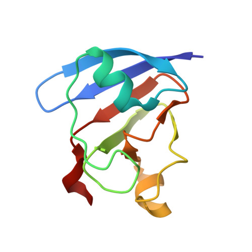
3P63 - PubMed Abstract:
Regulation of protein function via cracking, or local unfolding and refolding of substructures, is becoming a widely recognized mechanism of functional control. Oftentimes, cracking events are localized to secondary and tertiary structure interactions between domains that control the optimal position for catalysis and/or the formation of protein complexes. Small changes in free energy associated with ligand binding, phosphorylation, etc., can tip the balance and provide a regulatory functional switch. However, understanding the factors controlling function in single-domain proteins is still a significant challenge to structural biologists. We investigated the functional landscape of a single-domain plant-type ferredoxin protein and the effect of a distal loop on the electron-transfer center. We find the global stability and structure are minimally perturbed with mutation, whereas the functional properties are altered. Specifically, truncating the L1,2 loop does not lead to large-scale changes in the structure, determined via X-ray crystallography. Further, the overall thermal stability of the protein is only marginally perturbed by the mutation. However, even though the mutation is distal to the iron-sulfur cluster (∼20 Å), it leads to a significant change in the redox potential of the iron-sulfur cluster (57 mV). Structure-based all-atom simulations indicate correlated dynamical changes between the surface-exposed loop and the iron-sulfur cluster-binding region. Our results suggest intrinsic communication channels within the ferredoxin fold, composed of many short-range interactions, lead to the propagation of long-range signals. Accordingly, protein interface interactions that involve L1,2 could potentially signal functional changes in distal regions, similar to what is observed in other allosteric systems.
- Life Science Institute and The Wolfson Centre for Applied Structural Biology, The Hebrew University of Jerusalem, Edmond J. Safra Campus, Givat Ram, Jerusalem 91904, Israel.
Organizational Affiliation: