3ORM

Mycobacterium tuberculosis PknB kinase domain D76A mutant


Experimental Data Snapshot

  • Method: X-RAY DIFFRACTION
  • Resolution: 2.50 Å
  • R-Value Free: 0.288 
  • R-Value Work: 0.215 
  • R-Value Observed: 0.218 

wwPDB Validation   3D Report Full Report


Ligand Structure Quality Assessment 


This is version 1.3 of the entry. See complete history


Literature

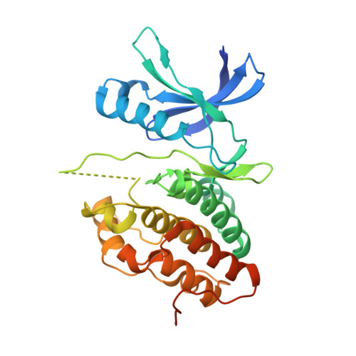
Allosteric activation mechanism of the Mycobacterium tuberculosis receptor Ser/Thr protein kinase, PknB.

Lombana, T.N.Echols, N.Good, M.C.Thomsen, N.D.Ng, H.L.Greenstein, A.E.Falick, A.M.King, D.S.Alber, T.

(2010) Structure 18: 1667-1677

  • DOI: https://doi.org/10.1016/j.str.2010.09.019
  • Primary Citation of Related Structures:  
    3ORI, 3ORM, 3ORO

  • PubMed Abstract: 

    The essential Mycobacterium tuberculosis Ser/Thr protein kinase (STPK), PknB, plays a key role in regulating growth and division, but the structural basis of activation has not been defined. Here, we provide biochemical and structural evidence that dimerization through the kinase-domain (KD) N-lobe activates PknB by an allosteric mechanism. Promoting KD pairing using a small-molecule dimerizer stimulates the unphosphorylated kinase, and substitutions that disrupt N-lobe pairing decrease phosphorylation activity in vitro and in vivo. Multiple crystal structures of two monomeric PknB KD mutants in complex with nucleotide reveal diverse inactive conformations that contain large active-site distortions that propagate > 30 Å from the mutation site. These results define flexible, inactive structures of a monomeric bacterial receptor KD and show how "back-to-back" N-lobe dimerization stabilizes the active KD conformation. This general mechanism of bacterial receptor STPK activation affords insights into the regulation of homologous eukaryotic kinases that form structurally similar dimers.


  • Organizational Affiliation

    Department of Molecular and Cell Biology, QB3 Institute, University of California, Berkeley, CA 94720-3200, USA.


Macromolecules
Find similar proteins by:  (by identity cutoff)  |  3D Structure
Entity ID: 1
MoleculeChains Sequence LengthOrganismDetailsImage
Serine/threonine protein kinase311Mycobacterium tuberculosis H37RaMutation(s): 1 
Gene Names: MRA_0016pknBPknB (Rv0014c)
UniProt
Find proteins for P9WI81 (Mycobacterium tuberculosis (strain ATCC 25618 / H37Rv))
Explore P9WI81 
Go to UniProtKB:  P9WI81
Entity Groups  
Sequence Clusters30% Identity50% Identity70% Identity90% Identity95% Identity100% Identity
UniProt GroupP9WI81
Sequence Annotations
Expand
  • Reference Sequence
Small Molecules
Ligands 2 Unique
IDChains Name / Formula / InChI Key2D Diagram3D Interactions
AGS
Query on AGS

Download Ideal Coordinates CCD File 
B [auth A]PHOSPHOTHIOPHOSPHORIC ACID-ADENYLATE ESTER
C10 H16 N5 O12 P3 S
NLTUCYMLOPLUHL-KQYNXXCUSA-N
MN
Query on MN

Download Ideal Coordinates CCD File 
C [auth A]MANGANESE (II) ION
Mn
WAEMQWOKJMHJLA-UHFFFAOYSA-N
Experimental Data & Validation

Experimental Data

  • Method: X-RAY DIFFRACTION
  • Resolution: 2.50 Å
  • R-Value Free: 0.288 
  • R-Value Work: 0.215 
  • R-Value Observed: 0.218 
  • Space Group: P 21 21 21
Unit Cell:
Length ( Å )Angle ( ˚ )
a = 40.458α = 90
b = 51.076β = 90
c = 134.746γ = 90
Software Package:
Software NamePurpose
Blu-Icedata collection
MOLREPphasing
PHENIXrefinement
HKL-2000data reduction
SCALEPACKdata scaling

Structure Validation

View Full Validation Report


Ligand Structure Quality Assessment 


Entry History 

Deposition Data

Revision History  (Full details and data files)

  • Version 1.0: 2010-12-15
    Type: Initial release
  • Version 1.1: 2011-07-13
    Changes: Version format compliance
  • Version 1.2: 2013-02-06
    Changes: Data collection, Database references, Refinement description
  • Version 1.3: 2023-09-06
    Changes: Data collection, Database references, Derived calculations, Refinement description