3OQG

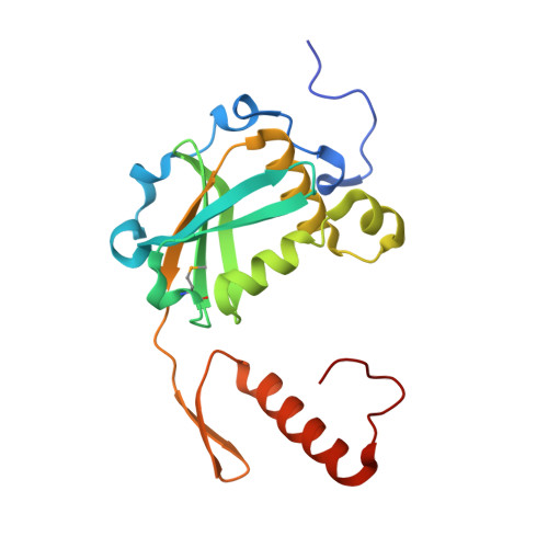
Restriction endonuclease HPY188I in complex with substrate DNA


Experimental Data Snapshot

  • Method: X-RAY DIFFRACTION
  • Resolution: 1.75 Å
  • R-Value Free: 0.192 
  • R-Value Work: 0.165 
  • R-Value Observed: 0.166 

wwPDB Validation   3D Report Full Report


This is version 1.2 of the entry. See complete history


Literature

Hpy188I-DNA pre- and post-cleavage complexes--snapshots of the GIY-YIG nuclease mediated catalysis.

Sokolowska, M.Czapinska, H.Bochtler, M.

(2011) Nucleic Acids Res 39: 1554-1564

  • DOI: https://doi.org/10.1093/nar/gkq821
  • Primary Citation of Related Structures:  
    3OQG, 3OR3

  • PubMed Abstract: 

    The GIY-YIG nuclease domain is present in all kingdoms of life and has diverse functions. It is found in the eukaryotic flap endonuclease and Holliday junction resolvase Slx1-Slx4, the prokaryotic nucleotide excision repair proteins UvrC and Cho, and in proteins of 'selfish' genetic elements. Here we present the structures of the ternary pre- and post-cleavage complexes of the type II GIY-YIG restriction endonuclease Hpy188I with DNA and a surrogate or catalytic metal ion, respectively. Our structures suggest that GIY-YIG nucleases catalyze DNA hydrolysis by a single substitution reaction. They are consistent with a previous proposal that a tyrosine residue (which we expect to occur in its phenolate form) acts as a general base for the attacking water molecule. In contrast to the earlier proposal, our data identify the general base with the GIY and not the YIG tyrosine. A conserved glutamate residue (Glu149 provided in trans in Hpy188I) anchors a single metal cation in the active site. This metal ion contacts the phosphate proS oxygen atom and the leaving group 3'-oxygen atom, presumably to facilitate its departure. Taken together, our data reveal striking analogy in the absence of homology between GIY-YIG and ββα-Me nucleases.


  • Organizational Affiliation

    International Institute of Molecular and Cell Biology, Trojdena 4, 02-109 Warsaw, Poland.


Macromolecules

Find similar proteins by:  (by identity cutoff)  |  3D Structure
Entity ID: 1
MoleculeChains Sequence LengthOrganismDetailsImage
Hpy188I
A, B
180Helicobacter pylori 118Mutation(s): 0 
Gene Names: hpy188IR
UniProt
Find proteins for Q9KJ88 (Helicobacter pylori)
Explore Q9KJ88 
Go to UniProtKB:  Q9KJ88
Entity Groups  
Sequence Clusters30% Identity50% Identity70% Identity90% Identity95% Identity100% Identity
UniProt GroupQ9KJ88
Sequence Annotations
Expand
  • Reference Sequence

Find similar nucleic acids by:  Sequence   |   3D Structure  

Entity ID: 2
MoleculeChains LengthOrganismImage
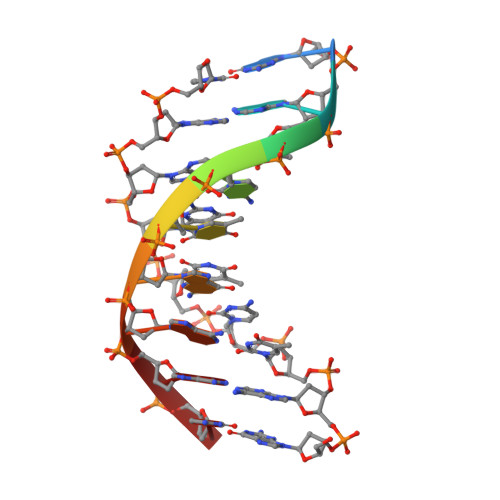
DNA 5'-D(*GP*AP*TP*CP*TP*GP*AP*AP*C)-3'9N/A
Sequence Annotations
Expand
  • Reference Sequence

Find similar nucleic acids by:  Sequence   |   3D Structure  

Entity ID: 3
MoleculeChains LengthOrganismImage
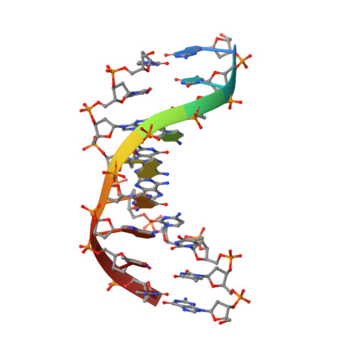
DNA 5'-D(*GP*TP*TP*CP*AP*GP*AP*TP*C)-3'9N/A
Sequence Annotations
Expand
  • Reference Sequence
Small Molecules
Modified Residues  1 Unique
IDChains TypeFormula2D DiagramParent
MSE
Query on MSE
A, B
L-PEPTIDE LINKINGC5 H11 N O2 SeMET
Experimental Data & Validation

Experimental Data

  • Method: X-RAY DIFFRACTION
  • Resolution: 1.75 Å
  • R-Value Free: 0.192 
  • R-Value Work: 0.165 
  • R-Value Observed: 0.166 
  • Space Group: P 41 21 2
Unit Cell:
Length ( Å )Angle ( ˚ )
a = 65.236α = 90
b = 65.236β = 90
c = 220.63γ = 90
Software Package:
Software NamePurpose
SHELXCDphasing
SHELXDphasing
SHELXEmodel building
MLPHAREphasing
DMmodel building
ARP/wARPmodel building
REFMACrefinement
DENZOdata reduction
SCALEPACKdata scaling
DMphasing

Structure Validation

View Full Validation Report


Entry History 

Deposition Data

Revision History  (Full details and data files)

  • Version 1.0: 2010-10-20
    Type: Initial release
  • Version 1.1: 2011-07-13
    Changes: Version format compliance
  • Version 1.2: 2017-11-08
    Changes: Refinement description