


Crystal Structure of Saccharomyces cerevisiae Dbf4-motif N
Matthews, L.A., Jones, D.R., Prasad, A.A., Duncker, B.P., Guarne, A.To be published.
Experimental Data Snapshot
Starting Model: experimental
View more details
wwPDB Validation 3D Report Full Report

Entity ID: 1 | |||||
|---|---|---|---|---|---|
| Molecule | Chains | Sequence Length | Organism | Details | Image |
| DBF4 | 134 | Saccharomyces cerevisiae | Mutation(s): 0 Gene Names: D4205, DBF4, DNA52, YD9609.07C, YDR052C | 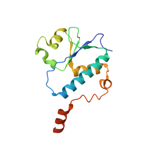 | |
UniProt | |||||
Entity Groups | |||||
| Sequence Clusters | 30% Identity50% Identity70% Identity90% Identity95% Identity100% Identity | ||||
| UniProt Group | P32325 | ||||
Sequence AnnotationsExpand | |||||
Reference Sequence | |||||
| Length ( Å ) | Angle ( ˚ ) |
|---|---|
| a = 83.714 | α = 90 |
| b = 99.707 | β = 90 |
| c = 127.045 | γ = 90 |
| Software Name | Purpose |
|---|---|
| CBASS | data collection |
| PHASER | phasing |
| PHENIX | refinement |
| HKL-2000 | data reduction |
| HKL-2000 | data scaling |


