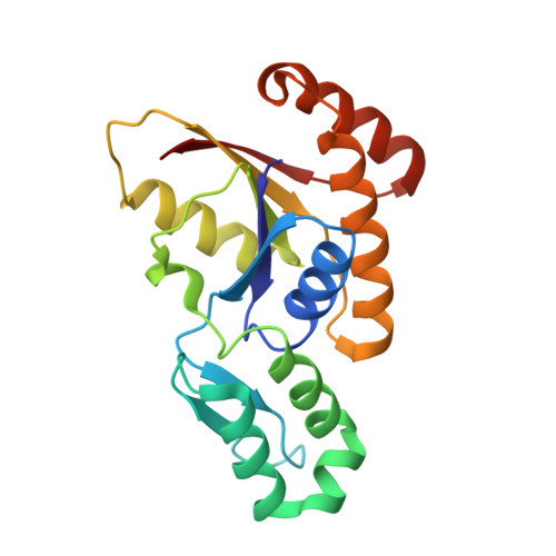
Crystal structure of Ssu72, an essential eukaryotic phosphatase specific for the C-terminal domain of RNA polymerase II, in complex with a transition state analogue.
Zhang, Y., Zhang, M., Zhang, Y.(2011) Biochem J 434: 435-444
- PubMed: 21204787 Search on PubMed
- DOI: https://doi.org/10.1042/BJ20101471
- Primary Citation Related Structures:
3OMW, 3OMX - PubMed Abstract:
Reversible phosphorylation of the CTD (C-terminal domain) of the eukaryotic RNA polymerase II largest subunit represents a critical regulatory mechanism during the transcription cycle and mRNA processing. Ssu72 is an essential phosphatase conserved in eukaryotes that dephosphorylates phosphorylated Ser5 of the CTD heptapeptide. Its function is implicated in transcription initiation, elongation and termination, as well as RNA processing. In the present paper we report the high resolution X-ray crystal structures of Drosophila melanogaster Ssu72 phosphatase in the apo form and in complex with an inhibitor mimicking the transition state of phosphoryl transfer. Ssu72 facilitates dephosphorylation of the substrate through a phosphoryl-enzyme intermediate, as visualized in the complex structure of Ssu72 with the oxo-anion compound inhibitor vanadate at a 2.35 Å (1 Å=0.1 nm) resolution. The structure resembles the transition state of the phosphoryl transfer with vanadate exhibiting a trigonal bi-pyramidal geometry covalently bonded to the nucleophilic cysteine residue. Interestingly, the incorporation of oxo-anion compounds greatly stabilizes a flexible loop containing the general acid, as detected by an increase of melting temperature of Ssu72 detected by differential scanning fluorimetry. The Ssu72 structure exhibits a core fold with a similar topology to that of LMWPTPs [low-molecular-mass PTPs (protein tyrosine phosphatases)], but with an insertion of a unique 'cap' domain to shelter the active site from the solvent with a deep groove in between where the CTD substrates bind. Mutagenesis studies in this groove established the functional roles of five residues (Met17, Pro46, Asp51, Tyr77 and Met85) that are essential specifically for substrate recognition.
- Department of Chemistry and Biochemistry, University of Texas at Austin, Austin, TX 78712, USA.
Organizational Affiliation: