3OJW | pdb_00003ojw

Disulfide crosslinked cytochrome P450 reductase inactive


Experimental Data Snapshot

  • Method: X-RAY DIFFRACTION
  • Resolution: 2.20 Å
  • R-Value Free: 
    0.251 (Depositor), 0.251 (DCC) 
  • R-Value Work: 
    0.205 (Depositor), 0.205 (DCC) 
  • R-Value Observed: 
    0.205 (Depositor) 

Starting Model: experimental
View more details

wwPDB Validation 3D Report Full Report

Validation slider image for 3OJW

Ligand Structure Quality Assessment 


This is version 1.4 of the entry. See complete history

Literature

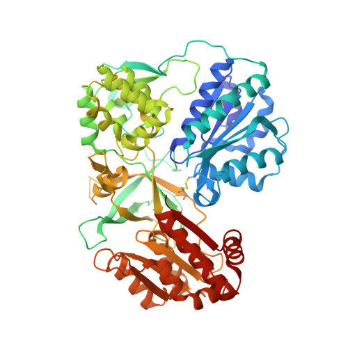
Conformational Changes of NADPH-Cytochrome P450 Oxidoreductase Are Essential for Catalysis and Cofactor Binding.

Xia, C.Hamdane, D.Shen, A.L.Choi, V.Kasper, C.B.Pearl, N.M.Zhang, H.Im, S.C.Waskell, L.Kim, J.J.

(2011) J Biological Chem 286: 16246-16260

  • DOI: https://doi.org/10.1074/jbc.M111.230532
  • Primary Citation Related Structures: 
    3OJW, 3OJX

  • PubMed Abstract: 

    The crystal structure of NADPH-cytochrome P450 reductase (CYPOR) implies that a large domain movement is essential for electron transfer from NADPH via FAD and FMN to its redox partners. To test this hypothesis, a disulfide bond was engineered between residues Asp(147) and Arg(514) in the FMN and FAD domains, respectively. The cross-linked form of this mutant protein, designated 147CC514, exhibited a significant decrease in the rate of interflavin electron transfer and large (≥90%) decreases in rates of electron transfer to its redox partners, cytochrome c and cytochrome P450 2B4. Reduction of the disulfide bond restored the ability of the mutant to reduce its redox partners, demonstrating that a conformational change is essential for CYPOR function. The crystal structures of the mutant without and with NADP(+) revealed that the two flavin domains are joined by a disulfide linkage and that the relative orientations of the two flavin rings are twisted ∼20° compared with the wild type, decreasing the surface contact area between the two flavin rings. Comparison of the structures without and with NADP(+) shows movement of the Gly(631)-Asn(635) loop. In the NADP(+)-free structure, the loop adopts a conformation that sterically hinders NADP(H) binding. The structure with NADP(+) shows movement of the Gly(631)-Asn(635) loop to a position that permits NADP(H) binding. Furthermore, comparison of these mutant and wild type structures strongly suggests that the Gly(631)-Asn(635) loop movement controls NADPH binding and NADP(+) release; this loop movement in turn facilitates the flavin domain movement, allowing electron transfer from FMN to the CYPOR redox partners.


  • Organizational Affiliation
    • Department of Biochemistry, Medical College of Wisconsin, Milwaukee, Wisconsin 53226, USA.

Macromolecule Content 

  • Total Structure Weight: 71.77 kDa 
  • Atom Count: 5,165 
  • Modeled Residue Count: 609 
  • Deposited Residue Count: 622 
  • Unique protein chains: 1

Macromolecules

Find similar proteins by:|  3D Structure
Entity ID: 1
MoleculeChains  Sequence LengthOrganismDetailsImage
NADPH-cytochrome P450 reductase622Rattus norvegicusMutation(s): 8 
Gene Names: CYPORPor
EC: 1.6.2.4
UniProt
Find proteins for P00388 (Rattus norvegicus)
Explore P00388 
Go to UniProtKB:  P00388
Entity Groups
Sequence Clusters30% Identity50% Identity70% Identity90% Identity95% Identity100% Identity
UniProt GroupP00388
Sequence Annotations
Expand
Reference Sequence

Small Molecules

Ligands 2 Unique
IDChains Name / Formula / InChI Key2D Diagram3D Interactions
FAD

Query on FAD


Download:Ideal Coordinates CCD File
C [auth A]FLAVIN-ADENINE DINUCLEOTIDE
C27 H33 N9 O15 P2
VWWQXMAJTJZDQX-UYBVJOGSSA-N
FMN

Query on FMN


Download:Ideal Coordinates CCD File
B [auth A]FLAVIN MONONUCLEOTIDE
C17 H21 N4 O9 P
FVTCRASFADXXNN-SCRDCRAPSA-N

Experimental Data & Validation

Experimental Data

  • Method: X-RAY DIFFRACTION
  • Resolution: 2.20 Å
  • R-Value Free:  0.251 (Depositor), 0.251 (DCC) 
  • R-Value Work:  0.205 (Depositor), 0.205 (DCC) 
  • R-Value Observed: 0.205 (Depositor) 
Space Group: P 21 21 21
Unit Cell:
Length ( Å )Angle ( ˚ )
a = 65.803α = 90
b = 72.958β = 90
c = 138.208γ = 90
Software Package:
Software NamePurpose
HKL-2000data collection
PHASERphasing
CNSrefinement
HKL-2000data reduction
HKL-2000data scaling

Structure Validation

View Full Validation Report


Ligand Structure Quality Assessment 


Entry History 

Deposition Data

Revision History  (Full details and data files)

  • Version 1.0: 2011-02-23
    Type: Initial release
  • Version 1.1: 2011-07-13
    Changes: Version format compliance
  • Version 1.2: 2018-03-07
    Changes: Data collection
  • Version 1.3: 2023-09-06
    Changes: Data collection, Database references, Derived calculations, Refinement description
  • Version 1.4: 2024-11-27
    Changes: Structure summary