3OG4

The crystal structure of human interferon lambda 1 complexed with its high affinity receptor in space group P21212


Experimental Data Snapshot

  • Method: X-RAY DIFFRACTION
  • Resolution: 2.16 Å
  • R-Value Free: 0.240 
  • R-Value Work: 0.198 
  • R-Value Observed: 0.199 

wwPDB Validation   3D Report Full Report


Ligand Structure Quality Assessment 


This is version 2.1 of the entry. See complete history


Literature

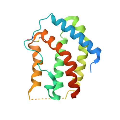
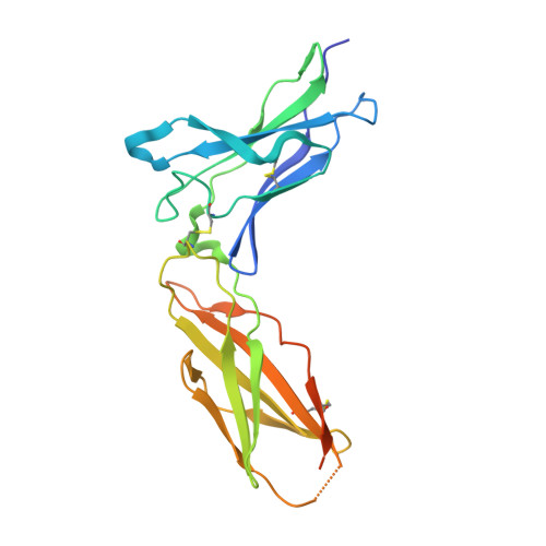
Crystal structure of the complex of human interferon-lambda1 with its high affinity receptor interferon-lambdaR1.

Miknis, Z.J.Magracheva, E.Li, W.Zdanov, A.Kotenko, S.V.Wlodawer, A.

(2010) J Mol Biol 404: 650-664

  • DOI: https://doi.org/10.1016/j.jmb.2010.09.068
  • Primary Citation of Related Structures:  
    3OG4, 3OG6

  • PubMed Abstract: 

    Interferon (IFN)-λ1 [also known as interleukin (IL)-29] belongs to the recently discovered group of type III IFNs. All type III IFNs initiate signaling processes through formation of specific heterodimeric receptor complexes consisting of IFN-λR1 and IL-10R2. We have determined the structure of human IFN-λ1 complexed with human IFN-λR1, a receptor unique to type III IFNs. The overall structure of IFN-λ1 is topologically similar to the structure of IL-10 and other members of the IL-10 family of cytokines. IFN-λR1 consists of two distinct domains having fibronectin type III topology. The ligand-receptor interface includes helix A, loop AB, and helix F on the IFN site, as well as loops primarily from the N-terminal domain and inter-domain hinge region of IFN-λR1. Composition and architecture of the interface that includes only a few direct hydrogen bonds support an idea that long-range ionic interactions between ligand and receptor govern the process of initial recognition of the molecules while hydrophobic interactions finalize it.


  • Organizational Affiliation

    Macromolecular Crystallography Laboratory, NCI-Frederick, Frederick, MD 21702, USA.


Macromolecules
Find similar proteins by:  (by identity cutoff)  |  3D Structure
Entity ID: 1
MoleculeChains Sequence LengthOrganismDetailsImage
Interleukin-29178Homo sapiensMutation(s): 0 
Gene Names: IFNL1IL29ZCYTO21
UniProt & NIH Common Fund Data Resources
Find proteins for Q8IU54 (Homo sapiens)
Explore Q8IU54 
Go to UniProtKB:  Q8IU54
PHAROS:  Q8IU54
GTEx:  ENSG00000182393 
Entity Groups  
Sequence Clusters30% Identity50% Identity70% Identity90% Identity95% Identity100% Identity
UniProt GroupQ8IU54
Sequence Annotations
Expand
  • Reference Sequence
Find similar proteins by:  (by identity cutoff)  |  3D Structure
Entity ID: 2
MoleculeChains Sequence LengthOrganismDetailsImage
Interleukin 28 receptor, alpha (Interferon, lambda receptor)226Homo sapiensMutation(s): 0 
Gene Names: hCG_1982865IL28RARP11-10N16.1-001
UniProt & NIH Common Fund Data Resources
Find proteins for Q8IU57 (Homo sapiens)
Explore Q8IU57 
Go to UniProtKB:  Q8IU57
PHAROS:  Q8IU57
GTEx:  ENSG00000185436 
Entity Groups  
Sequence Clusters30% Identity50% Identity70% Identity90% Identity95% Identity100% Identity
UniProt GroupQ8IU57
Sequence Annotations
Expand
  • Reference Sequence
Oligosaccharides

Help

Entity ID: 3
MoleculeChains Length2D Diagram Glycosylation3D Interactions
beta-D-mannopyranose-(1-3)-2-acetamido-2-deoxy-beta-D-glucopyranose-(1-4)-2-acetamido-2-deoxy-beta-D-glucopyranose
C
3N-Glycosylation
Glycosylation Resources
GlyTouCan:  G44266EX
GlyCosmos:  G44266EX
GlyGen:  G44266EX
Small Molecules
Ligands 1 Unique
IDChains Name / Formula / InChI Key2D Diagram3D Interactions
NAG
Query on NAG

Download Ideal Coordinates CCD File 
D [auth B]2-acetamido-2-deoxy-beta-D-glucopyranose
C8 H15 N O6
OVRNDRQMDRJTHS-FMDGEEDCSA-N
Experimental Data & Validation

Experimental Data

  • Method: X-RAY DIFFRACTION
  • Resolution: 2.16 Å
  • R-Value Free: 0.240 
  • R-Value Work: 0.198 
  • R-Value Observed: 0.199 
  • Space Group: P 21 21 2
Unit Cell:
Length ( Å )Angle ( ˚ )
a = 130.19α = 90
b = 65.44β = 90
c = 73.15γ = 90
Software Package:
Software NamePurpose
DENZOdata reduction
SCALEPACKdata scaling
PHASERphasing
PHENIXrefinement
PDB_EXTRACTdata extraction
HKL-2000data collection

Structure Validation

View Full Validation Report


Ligand Structure Quality Assessment 


Entry History 

Deposition Data

Revision History  (Full details and data files)

  • Version 1.0: 2010-10-20
    Type: Initial release
  • Version 1.1: 2011-07-13
    Changes: Version format compliance
  • Version 1.2: 2013-07-03
    Changes: Database references
  • Version 2.0: 2020-07-29
    Type: Remediation
    Reason: Carbohydrate remediation
    Changes: Advisory, Atomic model, Data collection, Database references, Derived calculations, Structure summary
  • Version 2.1: 2023-09-06
    Changes: Data collection, Database references, Refinement description, Structure summary