Compensating Stereochemical Changes Allow Murein Tripeptide to Be Accommodated in a Conventional Peptide-binding Protein.
Maqbool, A., Levdikov, V.M., Blagova, E.V., Herve, M., Horler, R.S., Wilkinson, A.J., Thomas, G.H.(2011) J Biological Chem 286: 31512-31521
- PubMed: 21705338 Search on PubMedSearch on PubMed Central
- DOI: https://doi.org/10.1074/jbc.M111.267179
- Primary Citation Related Structures:
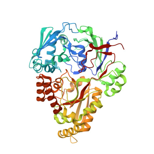
3O9P - PubMed Abstract:
The oligopeptide permease (Opp) of Escherichia coli is an ATP-binding cassette transporter that uses the substrate-binding protein (SBP) OppA to bind peptides and deliver them to the membrane components (OppBCDF) for transport. OppA binds conventional peptides 2-5 residues in length regardless of their sequence, but does not facilitate transport of the cell wall component murein tripeptide (Mtp, L-Ala-γ-D-Glu-meso-Dap), which contains a D-amino acid and a γ-peptide linkage. Instead, MppA, a homologous substrate-binding protein, forms a functional transporter with OppBCDF for uptake of this unusual tripeptide. Here we have purified MppA and demonstrated biochemically that it binds Mtp with high affinity (K(D) ∼ 250 nM). The crystal structure of MppA in complex with Mtp has revealed that Mtp is bound in a relatively extended conformation with its three carboxylates projecting from one side of the molecule and its two amino groups projecting from the opposite face. Specificity for Mtp is conferred by charge-charge and dipole-charge interactions with ionic and polar residues of MppA. Comparison of the structure of MppA-Mtp with structures of conventional tripeptides bound to OppA, reveals that the peptide ligands superimpose remarkably closely given the profound differences in their structures. Strikingly, the effect of the D-stereochemistry, which projects the side chain of the D-Glu residue at position 2 in the direction of the main chain in a conventional tripeptide, is compensated by the formation of a γ-linkage to the amino group of diaminopimelic acid, mimicking the peptide bond between residues 2 and 3 of a conventional tripeptide.
- Department of Biology (Area 10), University of York, York YO10 5YW, United Kingdom.
Organizational Affiliation: