3O8X

Recognition of Glycolipid Antigen by iNKT Cell TCR


Experimental Data Snapshot

  • Method: X-RAY DIFFRACTION
  • Resolution: 2.74 Å
  • R-Value Free: 0.253 
  • R-Value Work: 0.195 
  • R-Value Observed: 0.198 

wwPDB Validation   3D Report Full Report


Ligand Structure Quality Assessment 


This is version 2.2 of the entry. See complete history


Literature

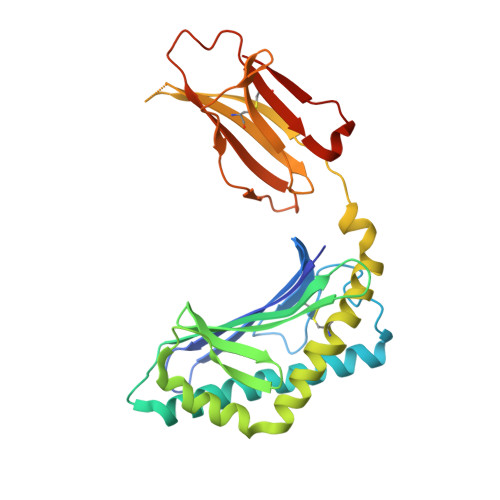
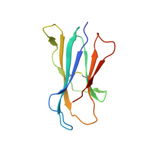
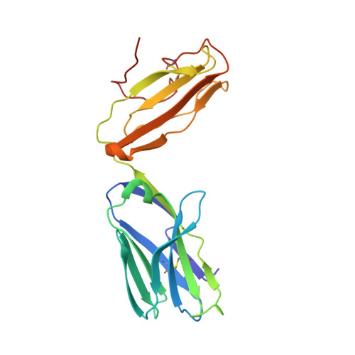
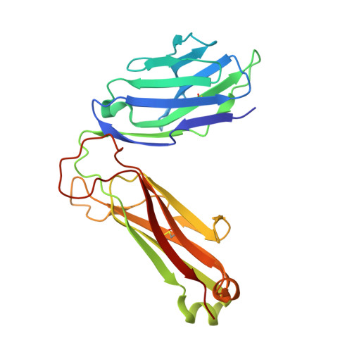
The V alpha 14 invariant natural killer T cell TCR forces microbial glycolipids and CD1d into a conserved binding mode.

Li, Y.Girardi, E.Wang, J.Yu, E.D.Painter, G.F.Kronenberg, M.Zajonc, D.M.

(2010) J Exp Med 207: 2383-2393

  • DOI: https://doi.org/10.1084/jem.20101335
  • Primary Citation of Related Structures:  
    3O8X, 3O9W

  • PubMed Abstract: 

    Invariant natural killer T cells (iNKT cells) rapidly produce effector cytokines. In this study, we report the first crystal structures of the iNKT cell T cell receptor (TCR) bound to two natural, microbial glycolipids presented by CD1d. Binding of the TCR induced CDR3-α-dependent structural changes in the F' roof of CD1d; these changes resemble those occurring in the absence of TCR engagement when the highly potent synthetic antigen α-galactosylceramide (α-GalCer) binds CD1d. Furthermore, in the Borrelia burgdorferi α-galactosyl diacylglycerol-CD1d complex, TCR binding caused a marked repositioning of the galactose sugar into an orientation that closely resembles α-GalCer. The TCR-dependent reorientation of the sugar, together with the induced CD1d fit, may explain the weaker potency of the microbial antigens compared with α-GalCer. We propose that the TCR of iNKT cells binds with a conserved footprint onto CD1d, regardless of the bound glycolipid antigen, and that for microbial antigens this unique binding mode requires TCR-initiated conformational changes.


  • Organizational Affiliation

    Division of Cell Biology, La Jolla Institute for Allergy and Immunology, La Jolla, CA 92037, USA.


Macromolecules
Find similar proteins by:  (by identity cutoff)  |  3D Structure
Entity ID: 1
MoleculeChains Sequence LengthOrganismDetailsImage
Antigen-presenting glycoprotein CD1d1285Mus musculusMutation(s): 1 
Gene Names: Cd1d1Cd1.1
UniProt & NIH Common Fund Data Resources
Find proteins for P11609 (Mus musculus)
Explore P11609 
Go to UniProtKB:  P11609
IMPC:  MGI:107674
Entity Groups  
Sequence Clusters30% Identity50% Identity70% Identity90% Identity95% Identity100% Identity
UniProt GroupP11609
Sequence Annotations
Expand
  • Reference Sequence
Find similar proteins by:  (by identity cutoff)  |  3D Structure
Entity ID: 2
MoleculeChains Sequence LengthOrganismDetailsImage
Beta-2-microglobulin99Mus musculusMutation(s): 0 
Gene Names: B2m
UniProt & NIH Common Fund Data Resources
Find proteins for P01887 (Mus musculus)
Explore P01887 
Go to UniProtKB:  P01887
IMPC:  MGI:88127
Entity Groups  
Sequence Clusters30% Identity50% Identity70% Identity90% Identity95% Identity100% Identity
UniProt GroupP01887
Sequence Annotations
Expand
  • Reference Sequence
Find similar proteins by:  (by identity cutoff)  |  3D Structure
Entity ID: 3
MoleculeChains Sequence LengthOrganismDetailsImage
Valpha14 chimera (Mouse variable domain, Human T-cell receptor alpha chain C region constant domain)209Mus musculusHomo sapiens
This entity is chimeric
Mutation(s): 1 
Gene Names: TRACTCRA
UniProt & NIH Common Fund Data Resources
Find proteins for P01848 (Homo sapiens)
Explore P01848 
Go to UniProtKB:  P01848
PHAROS:  P01848
Entity Groups  
Sequence Clusters30% Identity50% Identity70% Identity90% Identity95% Identity100% Identity
UniProt GroupP01848
Sequence Annotations
Expand
  • Reference Sequence
Find similar proteins by:  (by identity cutoff)  |  3D Structure
Entity ID: 4
MoleculeChains Sequence LengthOrganismDetailsImage
Vbeta8.2 chimera (Mouse variable domain, Human T-cell receptor beta-2 chain C region constant domain)241Mus musculusHomo sapiens
This entity is chimeric
Mutation(s): 2 
Gene Names: TRBC2TCRBC2
UniProt & NIH Common Fund Data Resources
Find proteins for A0A5B9 (Homo sapiens)
Explore A0A5B9 
Go to UniProtKB:  A0A5B9
PHAROS:  A0A5B9
Entity Groups  
Sequence Clusters30% Identity50% Identity70% Identity90% Identity95% Identity100% Identity
UniProt GroupA0A5B9
Sequence Annotations
Expand
  • Reference Sequence
Oligosaccharides

Help

Entity ID: 5
MoleculeChains Length2D Diagram Glycosylation3D Interactions
2-acetamido-2-deoxy-beta-D-glucopyranose-(1-4)-2-acetamido-2-deoxy-beta-D-glucopyranose
E
2N-Glycosylation
Glycosylation Resources
GlyTouCan:  G42666HT
GlyCosmos:  G42666HT
GlyGen:  G42666HT
Entity ID: 6
MoleculeChains Length2D Diagram Glycosylation3D Interactions
2-acetamido-2-deoxy-beta-D-glucopyranose-(1-4)-[alpha-L-fucopyranose-(1-6)]2-acetamido-2-deoxy-beta-D-glucopyranose
F
3N-Glycosylation
Glycosylation Resources
GlyTouCan:  G21290RB
GlyCosmos:  G21290RB
GlyGen:  G21290RB
Small Molecules
Ligands 2 Unique
IDChains Name / Formula / InChI Key2D Diagram3D Interactions
GSL
Query on GSL

Download Ideal Coordinates CCD File 
G [auth A](2S,3R)-3-HYDROXY-2-(TETRADECANOYLAMINO)OCTADECYL ALPHA-D-GALACTOPYRANOSIDURONIC ACID
C38 H73 N O9
IRPOZWRRAFKYMQ-LMIAXWKISA-N
NAG
Query on NAG

Download Ideal Coordinates CCD File 
H [auth A]2-acetamido-2-deoxy-beta-D-glucopyranose
C8 H15 N O6
OVRNDRQMDRJTHS-FMDGEEDCSA-N
Binding Affinity Annotations 
IDSourceBinding Affinity
TCR PDBBind:  3O8X Kd: 690 (nM) from 1 assay(s)
Experimental Data & Validation

Experimental Data

  • Method: X-RAY DIFFRACTION
  • Resolution: 2.74 Å
  • R-Value Free: 0.253 
  • R-Value Work: 0.195 
  • R-Value Observed: 0.198 
  • Space Group: C 2 2 21
Unit Cell:
Length ( Å )Angle ( ˚ )
a = 79α = 90
b = 191.2β = 90
c = 151.2γ = 90
Software Package:
Software NamePurpose
HKL-2000data collection
MOLREPphasing
REFMACrefinement
HKL-2000data reduction
HKL-2000data scaling

Structure Validation

View Full Validation Report


Ligand Structure Quality Assessment 


Entry History 

Deposition Data

Revision History  (Full details and data files)

  • Version 1.0: 2010-09-29
    Type: Initial release
  • Version 1.1: 2011-07-13
    Changes: Version format compliance
  • Version 1.2: 2017-06-21
    Changes: Database references, Source and taxonomy, Structure summary
  • Version 1.3: 2017-06-28
    Changes: Source and taxonomy
  • Version 2.0: 2020-07-29
    Type: Remediation
    Reason: Carbohydrate remediation
    Changes: Atomic model, Data collection, Derived calculations, Structure summary
  • Version 2.1: 2023-07-26
    Changes: Database references, Structure summary
  • Version 2.2: 2023-09-06
    Changes: Data collection, Refinement description