


Crystal Structures of apo-cyclophilin and bounded cyclosporine A from Moniliophthora perniciosa
Monzani, P.S., Pereira, H.M., Gramacho, K.P., Meirelles, F.V., Oliva, G., Cascardo, J.C.M.To be published.
Experimental Data Snapshot
wwPDB Validation 3D Report Full Report

Entity ID: 1 | |||||
|---|---|---|---|---|---|
| Molecule | Chains | Sequence Length | Organism | Details | Image |
| Cyclophilin A | 164 | Moniliophthora perniciosa | Mutation(s): 0 EC: 5.2.1.8 | 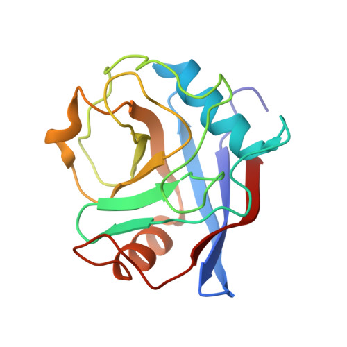 | |
UniProt | |||||
Entity Groups | |||||
| Sequence Clusters | 30% Identity50% Identity70% Identity90% Identity95% Identity100% Identity | ||||
| UniProt Group | E3P6K5 | ||||
Sequence AnnotationsExpand | |||||
Reference Sequence | |||||
| Length ( Å ) | Angle ( ˚ ) |
|---|---|
| a = 37.854 | α = 90 |
| b = 98.206 | β = 90 |
| c = 111.682 | γ = 90 |
| Software Name | Purpose |
|---|---|
| SCALA | data scaling |
| PHASER | phasing |
| PHENIX | refinement |
| PDB_EXTRACT | data extraction |
| MAR345dtb | data collection |
| MOSFLM | data reduction |


