3O4P | pdb_00003o4p

DFPase at 0.85 Angstrom resolution (H atoms included)


Experimental Data Snapshot

  • Method: X-RAY DIFFRACTION
  • Resolution: 0.85 Å
  • R-Value Free: 
    0.121 (Depositor), 0.124 (DCC) 
  • R-Value Work: 
    0.103 (Depositor), 0.110 (DCC) 

Starting Model: experimental
View more details

wwPDB Validation 3D Report Full Report

Validation slider image for 3O4P

This is version 1.3 of the entry. See complete history

Literature

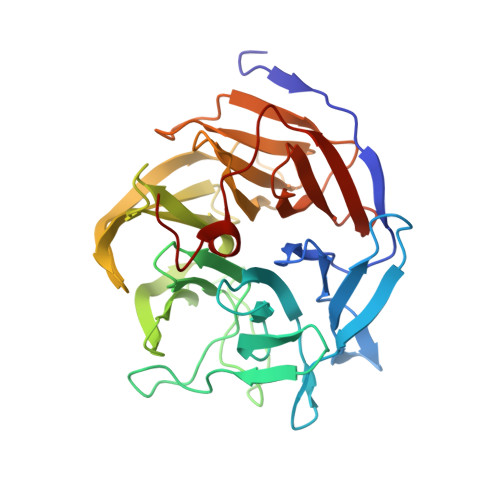
Hydrogen atoms in protein structures: high-resolution X-ray diffraction structure of the DFPase.

Elias, M.Liebschner, D.Koepke, J.Lecomte, C.Guillot, B.Jelsch, C.Chabriere, E.

(2013) BMC Res Notes 6: 308-308

  • DOI: https://doi.org/10.1186/1756-0500-6-308
  • Primary Citation Related Structures: 
    3O4P

  • PubMed Abstract: 

    Hydrogen atoms represent about half of the total number of atoms in proteins and are often involved in substrate recognition and catalysis. Unfortunately, X-ray protein crystallography at usual resolution fails to access directly their positioning, mainly because light atoms display weak contributions to diffraction. However, sub-Ångstrom diffraction data, careful modeling and a proper refinement strategy can allow the positioning of a significant part of hydrogen atoms. A comprehensive study on the X-ray structure of the diisopropyl-fluorophosphatase (DFPase) was performed, and the hydrogen atoms were modeled, including those of solvent molecules. This model was compared to the available neutron structure of DFPase, and differences in the protein and the active site solvation were noticed. A further examination of the DFPase X-ray structure provides substantial evidence about the presence of an activated water molecule that may constitute an interesting piece of information as regard to the enzymatic hydrolysis mechanism.


  • Organizational Affiliation
    • Weizmann Institute of Science, Biological Chemistry, Rehovot, Israel.

Macromolecule Content 

  • Total Structure Weight: 37.27 kDa 
  • Atom Count: 3,274 
  • Modeled Residue Count: 314 
  • Deposited Residue Count: 314 
  • Unique protein chains: 1

Macromolecules

Find similar proteins by:|  3D Structure
Entity ID: 1
MoleculeChains  Sequence LengthOrganismDetailsImage
Diisopropyl-fluorophosphatase314Loligo vulgarisMutation(s): 0 
EC: 3.1.8.2 (PDB Primary Data), 3.8.2.2 (UniProt)
UniProt
Find proteins for Q7SIG4 (Loligo vulgaris)
Explore Q7SIG4 
Go to UniProtKB:  Q7SIG4
Entity Groups
Sequence Clusters30% Identity50% Identity70% Identity90% Identity95% Identity100% Identity
UniProt GroupQ7SIG4
Sequence Annotations
Expand
Reference Sequence

Small Molecules

Ligands 9 Unique
IDChains Name / Formula / InChI Key2D Diagram3D Interactions
MES

Query on MES


Download:Ideal Coordinates CCD File
D [auth A],
E [auth A]
2-(N-MORPHOLINO)-ETHANESULFONIC ACID
C6 H13 N O4 S
SXGZJKUKBWWHRA-UHFFFAOYSA-N
PGE

Query on PGE


Download:Ideal Coordinates CCD File
N [auth A],
O [auth A]
TRIETHYLENE GLYCOL
C6 H14 O4
ZIBGPFATKBEMQZ-UHFFFAOYSA-N
ME2

Query on ME2


Download:Ideal Coordinates CCD File
V [auth A]1-ETHOXY-2-(2-METHOXYETHOXY)ETHANE
C7 H16 O3
CNJRPYFBORAQAU-UHFFFAOYSA-N
PEG

Query on PEG


Download:Ideal Coordinates CCD File
T [auth A],
U [auth A]
DI(HYDROXYETHYL)ETHER
C4 H10 O3
MTHSVFCYNBDYFN-UHFFFAOYSA-N
GOL

Query on GOL


Download:Ideal Coordinates CCD File
B [auth A],
C [auth A]
GLYCEROL
C3 H8 O3
PEDCQBHIVMGVHV-UHFFFAOYSA-N
DXE

Query on DXE


Download:Ideal Coordinates CCD File
P [auth A],
Q [auth A]
1,2-DIMETHOXYETHANE
C4 H10 O2
XTHFKEDIFFGKHM-UHFFFAOYSA-N
MXE

Query on MXE


Download:Ideal Coordinates CCD File
R [auth A],
S [auth A]
2-METHOXYETHANOL
C3 H8 O2
XNWFRZJHXBZDAG-UHFFFAOYSA-N
EDO

Query on EDO


Download:Ideal Coordinates CCD File
F [auth A]
G [auth A]
H [auth A]
I [auth A]
J [auth A]
F [auth A],
G [auth A],
H [auth A],
I [auth A],
J [auth A],
K [auth A],
L [auth A],
M [auth A]
1,2-ETHANEDIOL
C2 H6 O2
LYCAIKOWRPUZTN-UHFFFAOYSA-N
CA

Query on CA


Download:Ideal Coordinates CCD File
W [auth A],
X [auth A]
CALCIUM ION
Ca
BHPQYMZQTOCNFJ-UHFFFAOYSA-N

Experimental Data & Validation

Experimental Data

  • Method: X-RAY DIFFRACTION
  • Resolution: 0.85 Å
  • R-Value Free:  0.121 (Depositor), 0.124 (DCC) 
  • R-Value Work:  0.103 (Depositor), 0.110 (DCC) 
Space Group: P 21 21 21
Unit Cell:
Length ( Å )Angle ( ˚ )
a = 43.114α = 90
b = 81.849β = 90
c = 86.467γ = 90
Software Package:
Software NamePurpose
MoProrefinement
MOSFLMdata reduction
SCALAdata scaling

Structure Validation

View Full Validation Report


Entry History 

Deposition Data

Revision History  (Full details and data files)

  • Version 1.0: 2011-08-17
    Type: Initial release
  • Version 1.1: 2013-08-14
    Changes: Database references
  • Version 1.2: 2024-02-21
    Changes: Data collection, Database references, Derived calculations
  • Version 1.3: 2024-04-03
    Changes: Refinement description