3O1I

Crystal Structure of the TorS sensor domain - TorT complex in the absence of ligand


Experimental Data Snapshot

  • Method: X-RAY DIFFRACTION
  • Resolution: 2.80 Å
  • R-Value Free: 0.291 
  • R-Value Work: 0.248 
  • R-Value Observed: 0.248 

wwPDB Validation   3D Report Full Report


This is version 1.3 of the entry. See complete history


Literature

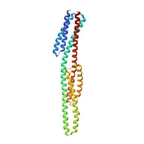
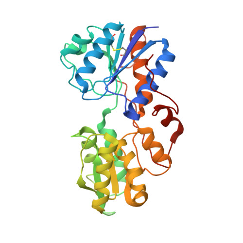
An asymmetry-to-symmetry switch in signal transmission by the histidine kinase receptor for TMAO.

Moore, J.O.Hendrickson, W.A.

(2012) Structure 20: 729-741

  • DOI: https://doi.org/10.1016/j.str.2012.02.021
  • Primary Citation of Related Structures:  
    3O1H, 3O1I, 3O1J

  • PubMed Abstract: 

    The osmoregulator trimethylamine-N-oxide (TMAO), commonplace in aquatic organisms, is used as the terminal electron acceptor for respiration in many bacterial species. The TMAO reductase (Tor) pathway for respiratory catalysis is controlled by a receptor system that comprises the TMAO-binding protein TorT, the sensor histidine kinase TorS, and the response regulator TorR. Here we study the TorS/TorT sensor system to gain mechanistic insight into signaling by histidine kinase receptors. We determined crystal structures for complexes of TorS sensor domains with apo TorT and with TorT (TMAO); we characterized TorS sensor associations with TorT in solution; we analyzed the thermodynamics of TMAO binding to TorT-TorS complexes; and we analyzed in vivo responses to TMAO through the TorT/TorS/TorR system to test structure-inspired hypotheses. TorS-TorT(apo) is an asymmetric 2:2 complex that binds TMAO with negative cooperativity to form a symmetric active kinase.


  • Organizational Affiliation

    Department of Biochemistry and Molecular Biophysics, Columbia University, New York, NY 10032, USA.


Macromolecules
Find similar proteins by:  (by identity cutoff)  |  3D Structure
Entity ID: 1
MoleculeChains Sequence LengthOrganismDetailsImage
Sensor protein TorS
A, B
277Vibrio parahaemolyticusMutation(s): 0 
Gene Names: TorSVPA0675
EC: 2.7.13.3
UniProt
Find proteins for Q87ID1 (Vibrio parahaemolyticus serotype O3:K6 (strain RIMD 2210633))
Explore Q87ID1 
Go to UniProtKB:  Q87ID1
Entity Groups  
Sequence Clusters30% Identity50% Identity70% Identity90% Identity95% Identity100% Identity
UniProt GroupQ87ID1
Sequence Annotations
Expand
  • Reference Sequence
Find similar proteins by:  (by identity cutoff)  |  3D Structure
Entity ID: 2
MoleculeChains Sequence LengthOrganismDetailsImage
Periplasmic protein TorTC [auth D],
D [auth C]
304Vibrio parahaemolyticusMutation(s): 0 
Gene Names: TorTVPA0674
UniProt
Find proteins for Q87ID2 (Vibrio parahaemolyticus serotype O3:K6 (strain RIMD 2210633))
Explore Q87ID2 
Go to UniProtKB:  Q87ID2
Entity Groups  
Sequence Clusters30% Identity50% Identity70% Identity90% Identity95% Identity100% Identity
UniProt GroupQ87ID2
Sequence Annotations
Expand
  • Reference Sequence
Small Molecules
Ligands 1 Unique
IDChains Name / Formula / InChI Key2D Diagram3D Interactions
PE4
Query on PE4

Download Ideal Coordinates CCD File 
E [auth B]2-{2-[2-(2-{2-[2-(2-ETHOXY-ETHOXY)-ETHOXY]-ETHOXY}-ETHOXY)-ETHOXY]-ETHOXY}-ETHANOL
C16 H34 O8
PJWQOENWHPEPKI-UHFFFAOYSA-N
Experimental Data & Validation

Experimental Data

  • Method: X-RAY DIFFRACTION
  • Resolution: 2.80 Å
  • R-Value Free: 0.291 
  • R-Value Work: 0.248 
  • R-Value Observed: 0.248 
  • Space Group: C 2 2 21
Unit Cell:
Length ( Å )Angle ( ˚ )
a = 115.71α = 90
b = 364.52β = 90
c = 80.81γ = 90
Software Package:
Software NamePurpose
SOLVEphasing
CNSrefinement
HKL-2000data reduction
SCALEPACKdata scaling

Structure Validation

View Full Validation Report


Entry History 

Deposition Data

Revision History  (Full details and data files)

  • Version 1.0: 2011-12-21
    Type: Initial release
  • Version 1.1: 2012-12-26
    Changes: Database references
  • Version 1.2: 2013-03-27
    Changes: Other
  • Version 1.3: 2024-04-03
    Changes: Data collection, Database references, Derived calculations, Refinement description