3NTC | pdb_00003ntc

Crystal structure of KD-247 Fab, an anti-V3 antibody that inhibits HIV-1 Entry


Experimental Data Snapshot

  • Method: X-RAY DIFFRACTION
  • Resolution: 1.55 Å
  • R-Value Free: 
    0.222 (Depositor), 0.208 (DCC) 
  • R-Value Work: 
    0.182 (Depositor), 0.168 (DCC) 
  • R-Value Observed: 
    0.184 (Depositor) 

Starting Model: experimental
View more details

wwPDB Validation 3D Report Full Report

Validation slider image for 3NTC

This is version 1.4 of the entry. See complete history

Literature

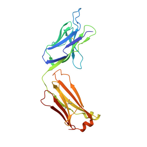
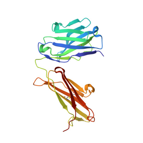
Structural basis of clade-specific HIV-1 neutralization by humanized anti-V3 monoclonal antibody KD-247.

Kirby, K.A.Ong, Y.T.Hachiya, A.Laughlin, T.G.Chiang, L.A.Pan, Y.Moran, J.L.Marchand, B.Singh, K.Gallazzi, F.Quinn, T.P.Yoshimura, K.Murakami, T.Matsushita, S.Sarafianos, S.G.

(2015) FASEB J 29: 70-80

  • DOI: https://doi.org/10.1096/fj.14-252262
  • Primary Citation Related Structures: 
    3NTC

  • PubMed Abstract: 

    Humanized monoclonal antibody KD-247 targets the Gly(312)-Pro(313)-Gly(314)-Arg(315) arch of the third hypervariable (V3) loop of the HIV-1 surface glycoprotein. It potently neutralizes many HIV-1 clade B isolates, but not of other clades. To understand the molecular basis of this specificity, we solved a high-resolution (1.55 Å) crystal structure of the KD-247 antigen binding fragment and examined the potential interactions with various V3 loop targets. Unlike most antibodies, KD-247 appears to interact with its target primarily through light chain residues. Several of these interactions involve Arg(315) of the V3 loop. To evaluate the role of light chain residues in the recognition of the V3 loop, we generated 20 variants of KD-247 single-chain variable fragments with mutations in the antigen-binding site. Purified proteins were assessed for V3 loop binding using AlphaScreen technology and for HIV-1 neutralization. Our data revealed that recognition of the clade-specificity defining residue Arg(315) of the V3 loop is based on a network of interactions that involve Tyr(L32), Tyr(L92), and Asn(L27d) that directly interact with Arg(315), thus elucidating the molecular interactions of KD-247 with its V3 loop target.


  • Organizational Affiliation
    • Christopher S. Bond Life Sciences Center, Department of Molecular Microbiology and Immunology, School of Medicine.

Macromolecule Content 

  • Total Structure Weight: 48.56 kDa 
  • Atom Count: 4,014 
  • Modeled Residue Count: 440 
  • Deposited Residue Count: 440 
  • Unique protein chains: 2

Macromolecules

Find similar proteins by:|  3D Structure
Entity ID: 1
MoleculeChains  Sequence LengthOrganismDetailsImage
Fab light chainA [auth L]219Homo sapiensMus musculus
This entity is chimeric
Mutation(s): 0 
Find similar proteins by:|  3D Structure
Entity ID: 2
MoleculeChains  Sequence LengthOrganismDetailsImage
Fab heavy chainB [auth H]221Homo sapiensMus musculus
This entity is chimeric
Mutation(s): 0 

Small Molecules

Ligands 3 Unique
IDChains Name / Formula / InChI Key2D Diagram3D Interactions
SO4

Query on SO4


Download:Ideal Coordinates CCD File
C [auth L],
J [auth H]
SULFATE ION
O4 S
QAOWNCQODCNURD-UHFFFAOYSA-L
EDO

Query on EDO


Download:Ideal Coordinates CCD File
D [auth L]
E [auth L]
F [auth L]
G [auth L]
H [auth L]
D [auth L],
E [auth L],
F [auth L],
G [auth L],
H [auth L],
I [auth L],
K [auth H],
L [auth H],
M [auth H],
N [auth H],
O [auth H],
P [auth H]
1,2-ETHANEDIOL
C2 H6 O2
LYCAIKOWRPUZTN-UHFFFAOYSA-N
ACT

Query on ACT


Download:Ideal Coordinates CCD File
Q [auth H]ACETATE ION
C2 H3 O2
QTBSBXVTEAMEQO-UHFFFAOYSA-M

Experimental Data & Validation

Experimental Data

  • Method: X-RAY DIFFRACTION
  • Resolution: 1.55 Å
  • R-Value Free:  0.222 (Depositor), 0.208 (DCC) 
  • R-Value Work:  0.182 (Depositor), 0.168 (DCC) 
  • R-Value Observed: 0.184 (Depositor) 
Space Group: P 21 21 21
Unit Cell:
Length ( Å )Angle ( ˚ )
a = 61.06α = 90
b = 69.18β = 90
c = 111.76γ = 90
Software Package:
Software NamePurpose
d*TREKdata scaling
MOLREPphasing
PHENIXrefinement
PDB_EXTRACTdata extraction
d*TREKdata reduction

Structure Validation

View Full Validation Report


Entry History 

Deposition Data

Revision History  (Full details and data files)

  • Version 1.0: 2010-08-25
    Type: Initial release
  • Version 1.1: 2011-07-13
    Changes: Version format compliance
  • Version 1.2: 2021-05-19
    Changes: Database references, Derived calculations
  • Version 1.3: 2023-09-06
    Changes: Data collection, Database references, Refinement description
  • Version 1.4: 2024-11-27
    Changes: Structure summary